Copyright
©The Author(s) 2005.
World J Gastroenterol. Aug 7, 2005; 11(29): 4478-4483
Published online Aug 7, 2005. doi: 10.3748/wjg.v11.i29.4478
Published online Aug 7, 2005. doi: 10.3748/wjg.v11.i29.4478
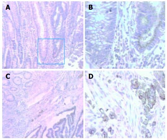
Figure 1 Immunohistochemical staining of mAb 3G11 in human colon carcinoma section.
A: Colon carcinoma (100×, the bar represents 400 μm in length.); B: the same case of A (400×, the bar represents 100 μm in length.); C (100×) and D (400×): another case of typical colon adenocarcinoma.
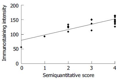
Figure 2 Immunohistochemical intensity of mAb 3G11 in various cases of human colorectal carcinoma assessed by image analysis and semiquantitative scoring.
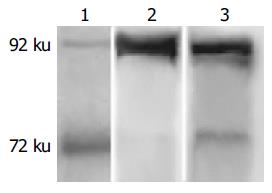
Figure 3 Immunological property of mAb 3G11 by Western blot analysis.
Lane 1: human colon cancer HT-29 cell lysate; Lane 2: human fibrosarcoma HT-1080 cell lysate; Lane 3: human breast cancer tissue lysate.
Figure 4 Gelatin-zymography assay of HT-29 cells after treatment with mAb 3G11 at different concentrations.
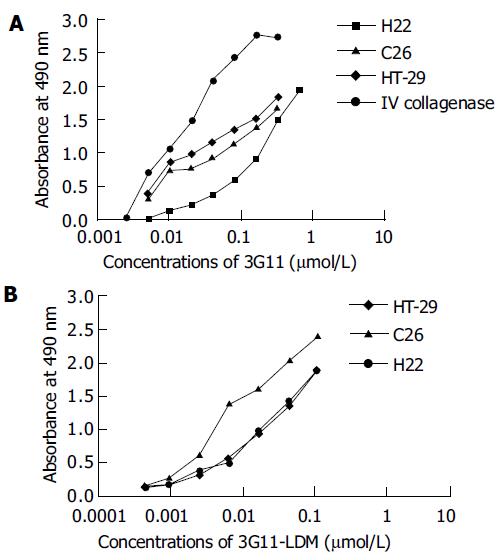
Figure 5 Immunoreactivity of 3G11(A) and 3G11-LDM (B) with type IV collagenase and various cancer cells in ELISA.
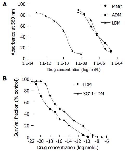
Figure 6 Cytotoxicity of LDM to human colon carcinoma HT-29 cells in comparison to ADM and MMC (A) and 3G11-LDM (B).
- Citation: Li L, Huang YH, Li Y, Wang FQ, Shang BY, Zhen YS. Antitumor activity of anti-type IV collagenase monoclonal antibody and its lidamycin conjugate against colon carcinoma. World J Gastroenterol 2005; 11(29): 4478-4483
- URL: https://www.wjgnet.com/1007-9327/full/v11/i29/4478.htm
- DOI: https://dx.doi.org/10.3748/wjg.v11.i29.4478


