Copyright
©The Author(s) 2005.
World J Gastroenterol. Jul 21, 2005; 11(27): 4261-4267
Published online Jul 21, 2005. doi: 10.3748/wjg.v11.i27.4261
Published online Jul 21, 2005. doi: 10.3748/wjg.v11.i27.4261
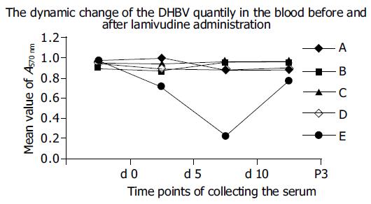
Figure 1 A: Lamivudine-susceptive DHBV control group; B: LRDHBV control group; C: LRDHBV-infected group administrated by large dose Lamivudine group; D: LRDHBV-infected group administrated by small dose Lamivudine group; E: Lamivudine-susceptive DHBV-infected group administrated by small dose Lamivudine group.
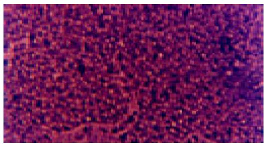
Figure 2 Duck embryo liver cells (5 d after DHBV inoculation, optic microscope × 40).
They have no significant effect of Duck embryo liver cells infected with DHBV or LRDHBV.
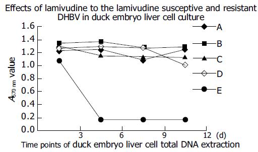
Figure 3 A: Lamivudine-susceptive DHBV control; B: LRDHBV control; C: LRDHBV-infected cells administrated by large dosage Lamivudine; D: LRDHBV-infected cells administrated by small dosage Lamivudine; E: Lamivudine-susceptive DHBV-infected cells administrated by small dosage Lamivudine.
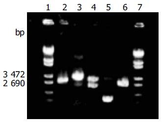
Figure 4 Electrophoresis graph of the amplification of DHBV complete genome and enzyme cutting of the recombinant.
Lanes 1 and 7: λ-EcoT14I digest marker; lane 2: the LAPCR product of DHBV complete genome; lane 3: the cloning product of DHBV complete genome; lane 4: EcoRI enzyme cutting product of DHBV complete genome recombinants; lane 5: loop product of pMD18-T vector; lane 6: EcoRI enzyme cutting product of loop product of pMD18-T vector.
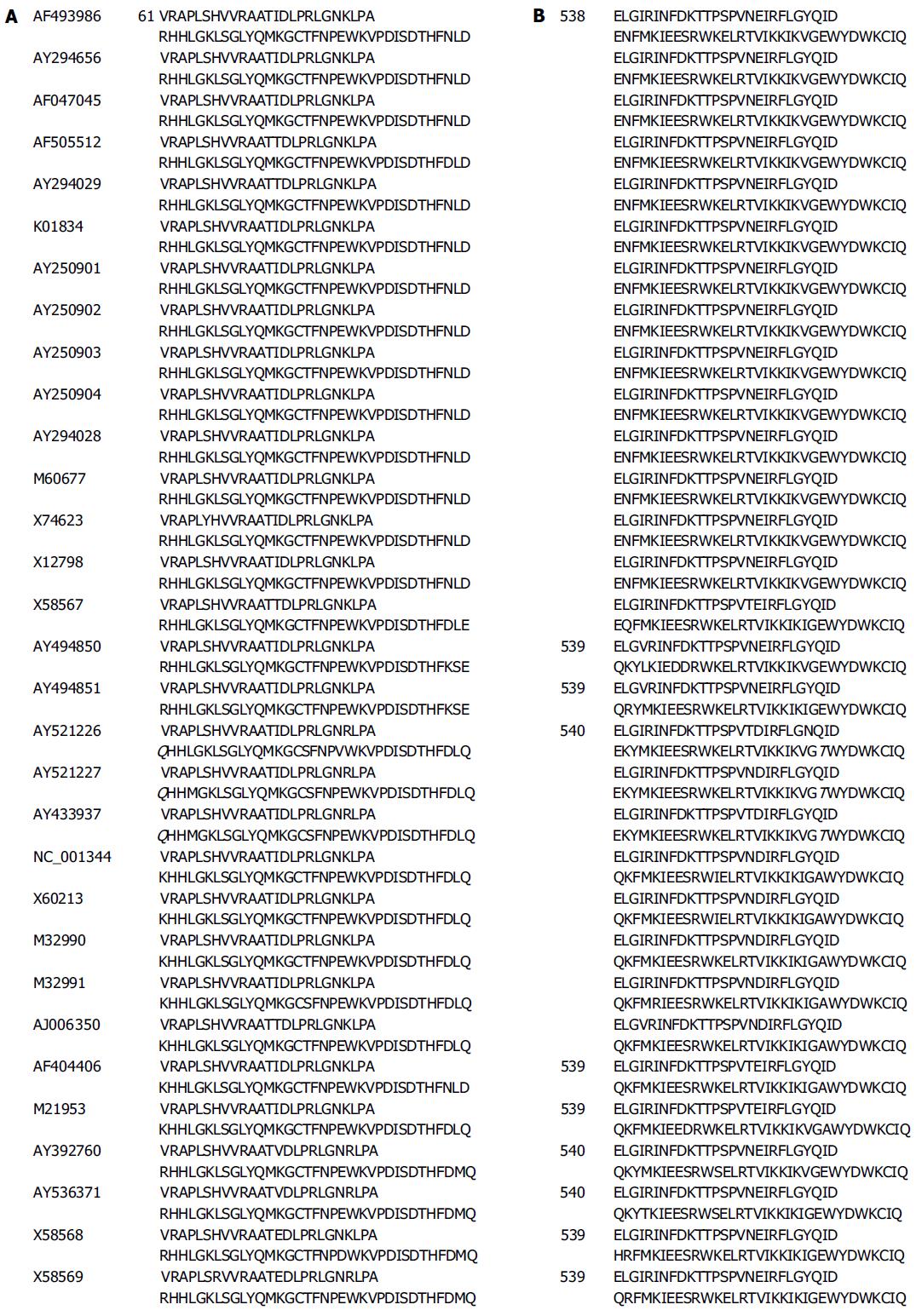
Figure 5 A: KorR86Q mutant point in the P protein of the LRDHBV; B: AorE591T mutant point in the P protein of the LRDHBV.
- Citation: He JY, Zhu YT, Yang RY, Feng LL, Guo XB, Zhang FX, Chen HS. Mutations outside the YMDD motif in the P protein can also cause DHBV resistant to Lamivudine. World J Gastroenterol 2005; 11(27): 4261-4267
- URL: https://www.wjgnet.com/1007-9327/full/v11/i27/4261.htm
- DOI: https://dx.doi.org/10.3748/wjg.v11.i27.4261


