Copyright
©The Author(s) 2005.
World J Gastroenterol. Jul 21, 2005; 11(27): 4140-4147
Published online Jul 21, 2005. doi: 10.3748/wjg.v11.i27.4140
Published online Jul 21, 2005. doi: 10.3748/wjg.v11.i27.4140
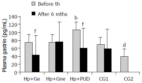
Figure 1 Basal plasma gastrin levels in patients with and without duodenal ulcer before and after eradication of H pylori infection.
H pylori+Ge-H pylori+eradicated patients with gastritis; H pylori+Gne-H pylori+non eradicated patients with gastritis; H pylori+DU- patients with H pylori+duodenal ulcer; CG1- H pylori negative dyspeptic patients; CG2- asymptomatic volunteers; th-therapy; mths-months; bP < 0.001 in H pylori+DU vs all other groups, dP < 0.01 in CG2 vs all other groups, fP < 0.001 before vs after eradication therapy.
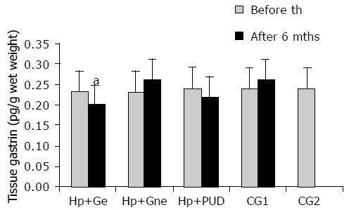
Figure 2 Antral tissue gastrin levels in patients with and without duodenal ulcer before and after eradication of H pylori infection.
H pylori +Ge-H pylori+ eradicated patients with gastritis; H pylori+Gne- H pylori+non eradicated patients with gastritis; H pylori +DU-patients with H pylori+duodenal ulcer; CG1- H pylori negative dyspeptic patients; CG2- asymptomatic volunteers; th-therapy; mths-months.
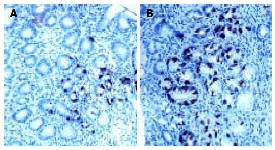
Figure 3 Pyloric gland area of H pylori associated gastritis before (A) and after eradication (B) therapy.
DAKO LSAB+ immunohistochemical staining and diaminobezydine (DAB) as chromogen; × 20 (A) and × 10 (B).There is evident lower gastrin cell number after succesful eradication therapy.
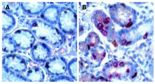
Figure 4 Synaptophysin and gastrin double immunostaining in pyloric gland of H pylori-associated gastritis before ( A) and after (B) eradication therapy.
DAKO EnVision double stain method; DAB is chromogen for G cells and aminoethylcarbazole (AEC) is chromogen for synaptophysin containing endocrine cells. × 20 (A) and × 10 ( B). G cells (red)/ other endocrine cells (brown) ratio is equal before (A) and after (B) succesful eradication of H pylori infection.
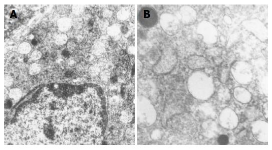
Figure 5 Electron micrographs of gastrin producing cells from healthy persons antral mucosa.
Normal ultrastructure is characterized by the presence of numerous secretory granules of different electron density. Uranyl acetate, lead citrate; × 8 400 (A) and × 12 680 (B).
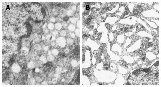
Figure 6 Electron micrographs of gastrin producing cells from H pylori infected individuals with gastritis (A) and duodenal ulcer ( B) before eradication therapy.
Uranyl acetate, lead citrate; x8 400 (both A and B). Note that total number of granules is higher than in controls and that dense core granules are more numerous in H pylori+patient with gastritis (A). Endoplasmatic reticulum is very prominent in H pylori+patient with duodenal ulcer.
-
Citation: Sokic-Milutinovic A, Todorovic V, Milosavljevic T, Micev M, Drndarevic N, Mitrovic O. Gastrin and antral G cells in course of
Helicobacter pylori eradication: Six months follow up study. World J Gastroenterol 2005; 11(27): 4140-4147 - URL: https://www.wjgnet.com/1007-9327/full/v11/i27/4140.htm
- DOI: https://dx.doi.org/10.3748/wjg.v11.i27.4140


