Copyright
©The Author(s) 2005.
World J Gastroenterol. Jul 7, 2005; 11(25): 3899-3904
Published online Jul 7, 2005. doi: 10.3748/wjg.v11.i25.3899
Published online Jul 7, 2005. doi: 10.3748/wjg.v11.i25.3899
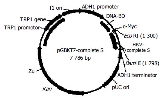
Figure 1 Structure of “bait” plasmid pGBKT7-complete S.
Figure 2 One thousand three hundred and thirty eight basepair fragment-complete S amplified by RT-PCR (A), pGBKT7-complete S cut by EcoRI/ BamHI (B), a 945 bp fragment-CSBP1 amplified by RT-PCR (C), pGBKT7-CSBP1 and pGADT7-CSBP1 cut by EcoRI/BamHI (D).
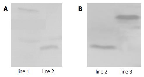
Figure 3 Expression of HBV complete S and HBV CSBP1 protein in yeast confirmed by Western blotting.
Lane 1: HBV complete S protein; lane 2: positive control; lane 3: HBV CSBP1 protein.
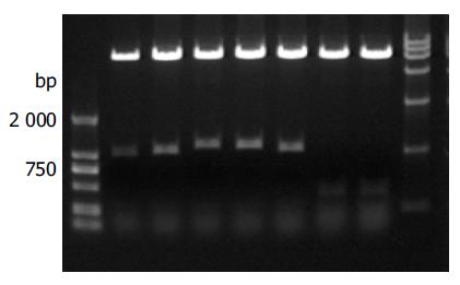
Figure 4 Identification of different colonies with BglII digestion.
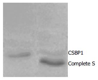
Figure 5 Interaction between HBV complete S protein and CSBP1 protein identified by coimmunoprecipitation.
Lane 1: HBV complete S protein; lane 2: interaction with two proteins.
- Citation: Bai GQ, Cheng J, Zhang SL, Huang YP, Wang L, Liu Y, Lin SM. Screening of hepatocyte proteins binding to complete S protein of hepatitis B virus by yeast-two hybrid system. World J Gastroenterol 2005; 11(25): 3899-3904
- URL: https://www.wjgnet.com/1007-9327/full/v11/i25/3899.htm
- DOI: https://dx.doi.org/10.3748/wjg.v11.i25.3899


