Copyright
©The Author(s) 2005.
World J Gastroenterol. Jul 7, 2005; 11(25): 3860-3865
Published online Jul 7, 2005. doi: 10.3748/wjg.v11.i25.3860
Published online Jul 7, 2005. doi: 10.3748/wjg.v11.i25.3860
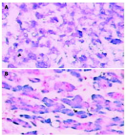
Figure 1 Hepatocellular carcinoma showing nuclear and cytoplasmic positivity positivity for NF-κB (A) and AP-1(B).
In situ hybridization (magnification ×200).
Figure 2 Nontumor liver tissue showing focal and weak cytoplasmic for NF-κB (A) and AP-1 (B).
In situ hybridization (magnification ×200).
Figure 3 Immunohistochemistry staining showing strong cytoplasmic positivity in adjacent hepatocytes and weak positivity in HCC for Fas (A), FasL (B), ICH- lL (C) and ICH-lS (D) (magnification ×200).
- Citation: Guo LL, Xiao S, Guo Y. Activation of transcription factors NF-kappaB and AP-1 and their relations with apoptosis-associated proteins in hepatocellular carcinoma. World J Gastroenterol 2005; 11(25): 3860-3865
- URL: https://www.wjgnet.com/1007-9327/full/v11/i25/3860.htm
- DOI: https://dx.doi.org/10.3748/wjg.v11.i25.3860


