Copyright
©The Author(s) 2005.
World J Gastroenterol. Jul 7, 2005; 11(25): 3855-3859
Published online Jul 7, 2005. doi: 10.3748/wjg.v11.i25.3855
Published online Jul 7, 2005. doi: 10.3748/wjg.v11.i25.3855
Figure 1 Expression of survivin in HCC and liver cirrhosis tissues (SP method, ×200).
A: The brown granules in the cytoplasm indicate survivin protein in liver cancer cells; B: Survivin protein is not detected in liver cirrhosis tissues.
Figure 2 Expression of VEGF in HCC and liver cirrhosis tissues (SP method, HCC; B: Expression of VEGF was weak positive in some liver cirrhosis ×100).
A: The brown granules in the cytoplasm indicate VEGF expression in tissues.
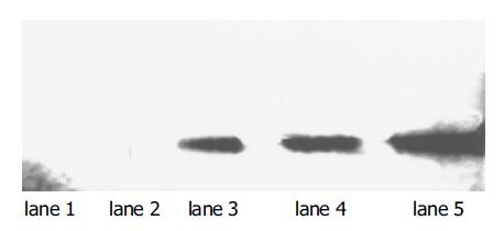
Figure 3 Expression of survivin in HCC and liver cirrhosis tissues.
Lanes 1 and 2: liver cirrhosis tissues; lanes 3-5: HCC tissues. Mr of survivin: 16500.
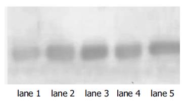
Figure 4 Expression of VEGF in HCC and liver cirrhosis tissues.
Lanes 1 and 2: liver cirrhosis tissues; lanes 3-5: HCC tissues. Mr of VEGF: 21000.
- Citation: Zhu H, Chen XP, Zhang WG, Luo SF, Zhang BX. Expression and significance of new inhibitor of apoptosis protein survivin in hepatocellular carcinoma. World J Gastroenterol 2005; 11(25): 3855-3859
- URL: https://www.wjgnet.com/1007-9327/full/v11/i25/3855.htm
- DOI: https://dx.doi.org/10.3748/wjg.v11.i25.3855


