Copyright
©2005 Baishideng Publishing Group Inc.
World J Gastroenterol. Jun 28, 2005; 11(24): 3797-3799
Published online Jun 28, 2005. doi: 10.3748/wjg.v11.i24.3797
Published online Jun 28, 2005. doi: 10.3748/wjg.v11.i24.3797
Figure 1 Computerized tomography scan of the abdomen.
(A) and (B) show the sagittal and coronal images of abdomen, respectively. Hepatomegaly with inhomogeneous distribution of contrast medium is visualized. Massive ascites is also recognized.
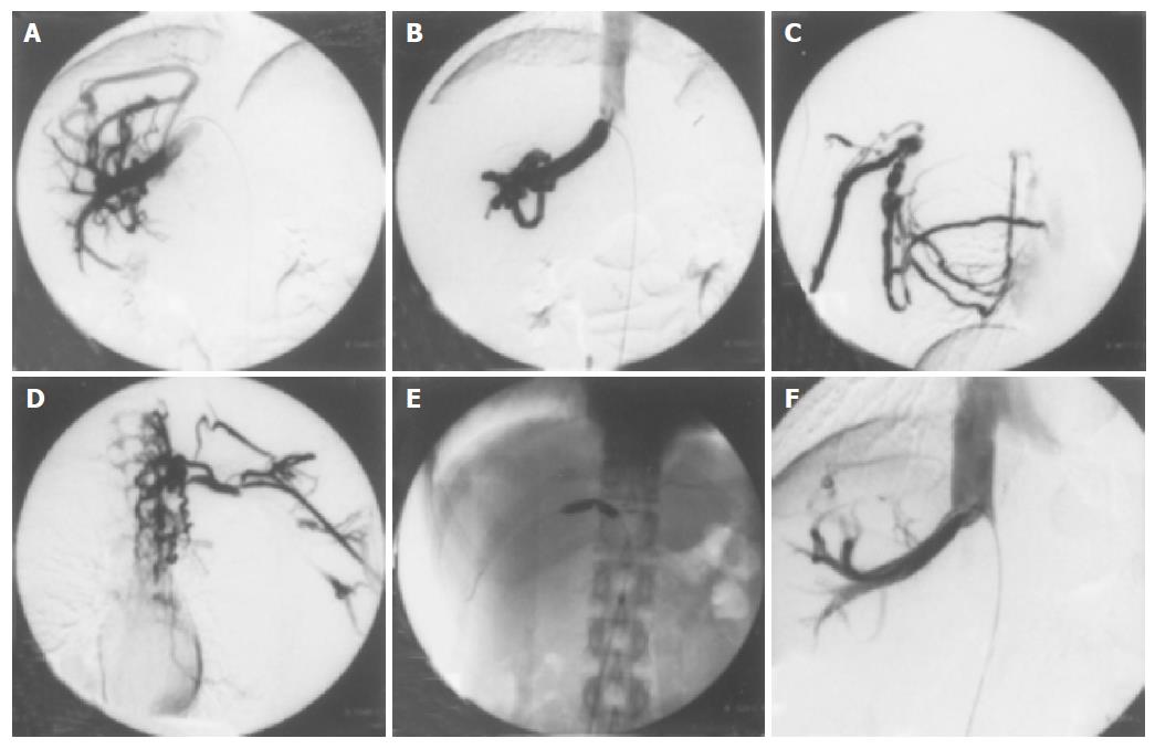
Figure 2 Cavography via the femoral vein and median basilic veins.
(A) and (B) show cavography via the femoral vein. Normal hepatic vein is not visualized and extensive collateral circulation (the right accessory hepatic vein) flows into the inferior vena cava. The orifice of this collateral vein is remarkably stenotic. (C) and (D) show cavography via right and left median basilic veins, respectively. Complete obstruction from the axillary to the subclavian vein and collateral circulation towards the right atrium are visualized. (E) shows a percutaneous balloon dilatation therapy for the stenosis in the orifice of her right accessory hepatic vein. (F) shows the dilated orifice of her right accessory hepatic vein after a balloon dilatation therapy.
- Citation: Araki Y, Sakaguchi C, Ishizuka I, Sasaki M, Tsujikawa T, Koyama S, Furukawa A, Fujiyama Y. Budd-Chiari syndrome: A case with a combination of hepatic vein and superior vena cava occlusion. World J Gastroenterol 2005; 11(24): 3797-3799
- URL: https://www.wjgnet.com/1007-9327/full/v11/i24/3797.htm
- DOI: https://dx.doi.org/10.3748/wjg.v11.i24.3797


