Copyright
©2005 Baishideng Publishing Group Inc.
World J Gastroenterol. Jun 28, 2005; 11(24): 3794-3796
Published online Jun 28, 2005. doi: 10.3748/wjg.v11.i24.3794
Published online Jun 28, 2005. doi: 10.3748/wjg.v11.i24.3794
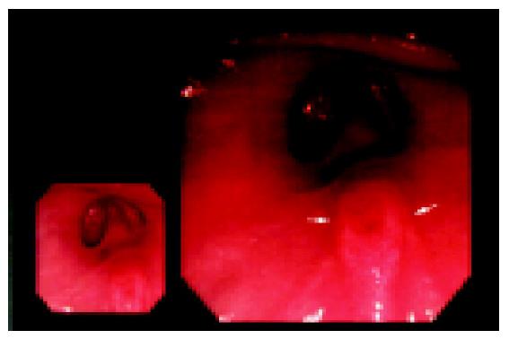
Figure 1 Swelling accessory papilla with ulcer (arrows) by endoscopy.
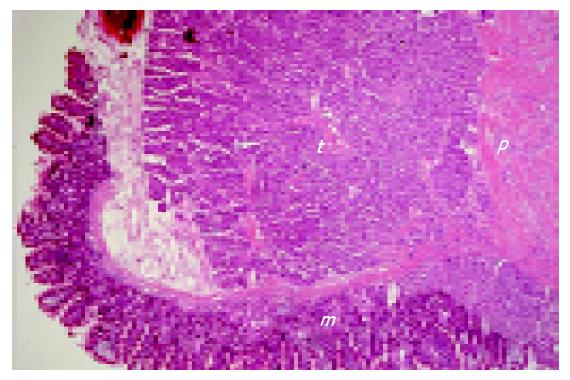
Figure 2 Tumor cells (t) in deep portion of duodenal mucosa (m) over the papilla and in submucosa around the ductal wall of papilla (p).
(H&E stain, ×20).
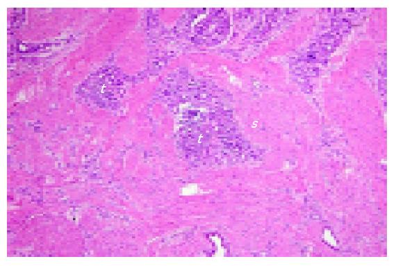
Figure 3 Tumor cells (t) with glandular pattern infiltrate the smooth muscle bundles of papilla (s).
(H&E stain, ×40).
- Citation: Wang HY, Chen MJ, Yang TL, Chang MC, Chan YJ. Carcinoid tumor of the duodenum and accessory papilla associated with polycythemia vera. World J Gastroenterol 2005; 11(24): 3794-3796
- URL: https://www.wjgnet.com/1007-9327/full/v11/i24/3794.htm
- DOI: https://dx.doi.org/10.3748/wjg.v11.i24.3794


