Copyright
©2005 Baishideng Publishing Group Inc.
World J Gastroenterol. Jun 28, 2005; 11(24): 3696-3700
Published online Jun 28, 2005. doi: 10.3748/wjg.v11.i24.3696
Published online Jun 28, 2005. doi: 10.3748/wjg.v11.i24.3696
Figure 1 Normal lobular architecture of mature and young liver (A) and normal distribution of collagen in mature and young liver (B).
Figure 2 Severe fatty degeneration (A) and collagen deposition (B) in aging liver.
Figure 3 Expression of TIMP-1 in mature and young liver (A) and aging liver (B).
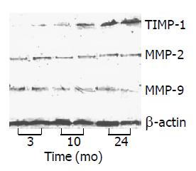
Figure 4 Protein expressions of TIMP-1 and MMP-2, MMP-9 in rat livers (Western blot).
- Citation: Zhang YM, Chen XM, Wu D, Shi SZ, Yin Z, Ding R, Lü Y. Expression of tissue inhibitor of matrix metalloproteinases-1 during aging in rat liver. World J Gastroenterol 2005; 11(24): 3696-3700
- URL: https://www.wjgnet.com/1007-9327/full/v11/i24/3696.htm
- DOI: https://dx.doi.org/10.3748/wjg.v11.i24.3696


