Copyright
©2005 Baishideng Publishing Group Inc.
World J Gastroenterol. Jun 14, 2005; 11(22): 3392-3397
Published online Jun 14, 2005. doi: 10.3748/wjg.v11.i22.3392
Published online Jun 14, 2005. doi: 10.3748/wjg.v11.i22.3392
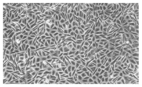
Figure 1 A confluent monolayer of KKU-100 cells shows compact polygonal to spindle cells.
(Phase-contrast, original magnification, ×100).
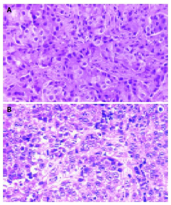
Figure 2 Histopathology of the primary tumor (A) and KKU-100 heterotransplanted tumors (B) in nude mice.
(hematoxylin and eosin, original magnification ×200).
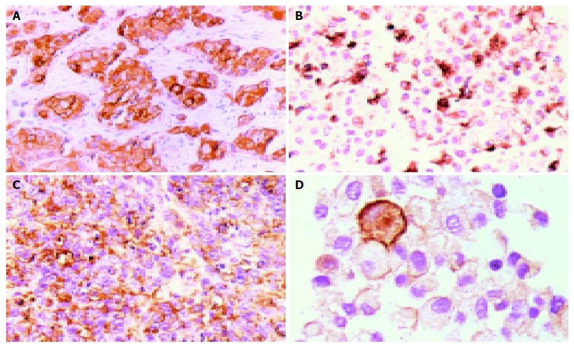
Figure 3 Immunocytochemistry of the primary tumor and KKU-100 cells.
A: the primary tumor showing strong expression of cytokeratin; B: strong expression of KKU-100 cell line; C: EMA expression in both heterotransplanted tumor cells; D: EMA expression in KKU-100 cells (immunoperoxidase, original magnification ×200).
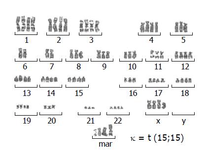
Figure 4 Representative G-banded karyotype of KKU-100 at passage 20 showing chromosomal abnormalities and marker chromosome (mar) : 84, +XX, +1, +2, +3, +4, +5, +8, +9, 10, +11, +12, +13, +14, +der(15)t(15;15), +16, +17, +18, +19, +20, +21, +22, +mar.
- Citation: Sripa B, Leungwattanawanit S, Nitta T, Wongkham C, Bhudhisawasdi V, Puapairoj A, Sripa C, Miwa M. Establishment and characterization of an opisthorchiasis-associated cholangiocarcinoma cell line (KKU-100). World J Gastroenterol 2005; 11(22): 3392-3397
- URL: https://www.wjgnet.com/1007-9327/full/v11/i22/3392.htm
- DOI: https://dx.doi.org/10.3748/wjg.v11.i22.3392


