Copyright
©2005 Baishideng Publishing Group Inc.
World J Gastroenterol. Jun 7, 2005; 11(21): 3311-3314
Published online Jun 7, 2005. doi: 10.3748/wjg.v11.i21.3311
Published online Jun 7, 2005. doi: 10.3748/wjg.v11.i21.3311
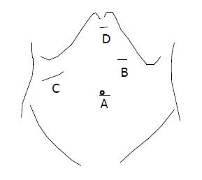
Figure 1 Position of the Trocars and the hand.
A: Camera port; B: working port; C: hand insertion; D: additional port.
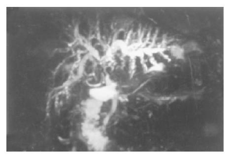
Figure 2 Preoperative MRCP demonstrated compression of the CHD by a large stone with dilation of intrahepatic bile duct characteristic of Mirizzi’s syndrome (Case 4).
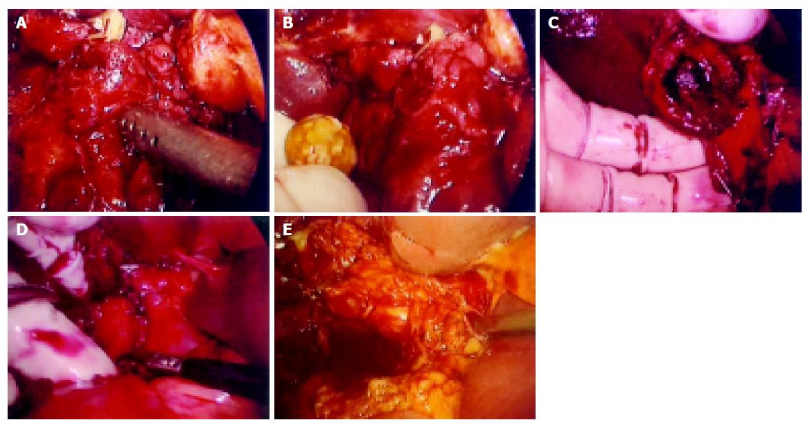
Figure 3 Causes of hand-assisted laparoscopic surgery.
A: One 1.0-cm stone impacted in the infundibulum fused with the CHD, impossible to remove with laparoscopic instruments; B: Using the intra-abdominal hand to facilitate such maneuvers as squeezing out of the stone (case 1); C: Compression of the CHD by one 2.5-cm stone; D: Identification and dissection of the obscured Calot triangle by the hand (case 4); E: A severely inflamed gallbladder with a neck stone caused obscured Calot’ triangle without jaundice (mimic Mirizzi syndrome, case 2).
- Citation: Wei Q, Shen LG, Zheng HM. Hand-assisted laparoscopic surgery for complex gallstone disease: A report of five cases. World J Gastroenterol 2005; 11(21): 3311-3314
- URL: https://www.wjgnet.com/1007-9327/full/v11/i21/3311.htm
- DOI: https://dx.doi.org/10.3748/wjg.v11.i21.3311


