Copyright
©2005 Baishideng Publishing Group Inc.
World J Gastroenterol. May 14, 2005; 11(18): 2792-2795
Published online May 14, 2005. doi: 10.3748/wjg.v11.i18.2792
Published online May 14, 2005. doi: 10.3748/wjg.v11.i18.2792
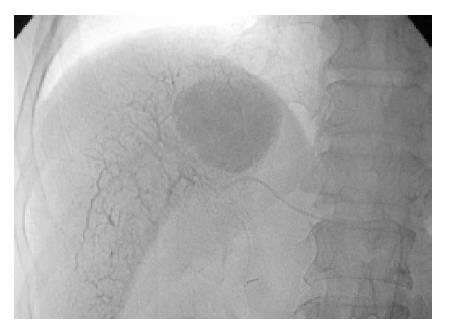
Figure 1 Post-embolization radiographic image of upper abdomen shows homogenously packed HCC and retrograde filling of multiple parenchymal portal veins.
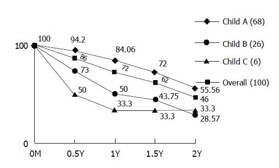
Figure 2 Survival rates of HCC in Child-Pugh classification.
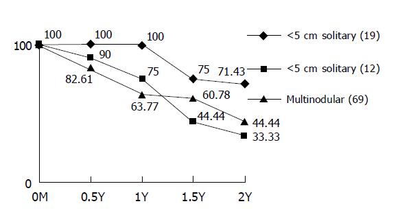
Figure 3 Survival rates of HCC in different morphology.
- Citation: Cheung YC, Ko SF, Ng SH, Chan SC, Cheng YF. Survival outcome of lobar or segmental transcatheter arterial embolization with ethanol-lipiodol mixture in treating hepatocellular carcinoma. World J Gastroenterol 2005; 11(18): 2792-2795
- URL: https://www.wjgnet.com/1007-9327/full/v11/i18/2792.htm
- DOI: https://dx.doi.org/10.3748/wjg.v11.i18.2792


