Copyright
©2005 Baishideng Publishing Group Inc.
World J Gastroenterol. May 14, 2005; 11(18): 2784-2788
Published online May 14, 2005. doi: 10.3748/wjg.v11.i18.2784
Published online May 14, 2005. doi: 10.3748/wjg.v11.i18.2784
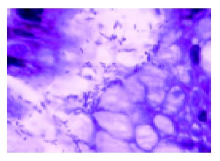
Figure 1 Photomicrography: antral biopsy (Fucsin stain, 1000×).
Presence of numerous (+++/+++) H pylori bacteria, forming clusters over the epithelial foveolar surface.
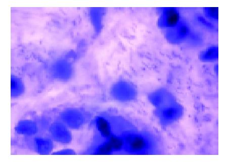
Figure 2 Photomicrography: antral exfoliative cytology (Papanicolaou stain, 1000×).
Presence of numerous (+++/+++) H pylori bacteria distributed among the epithelial cells, forming clusters.
-
Citation: Gomes Jr CA, Catapani WR, Mader AM, Locatelli Â, Silva CB, Waisberg J. Antral exfoliative cytology for the detection of
Helicobacter pylori in the stomach. World J Gastroenterol 2005; 11(18): 2784-2788 - URL: https://www.wjgnet.com/1007-9327/full/v11/i18/2784.htm
- DOI: https://dx.doi.org/10.3748/wjg.v11.i18.2784


