Copyright
©2005 Baishideng Publishing Group Inc.
World J Gastroenterol. May 14, 2005; 11(18): 2709-2713
Published online May 14, 2005. doi: 10.3748/wjg.v11.i18.2709
Published online May 14, 2005. doi: 10.3748/wjg.v11.i18.2709
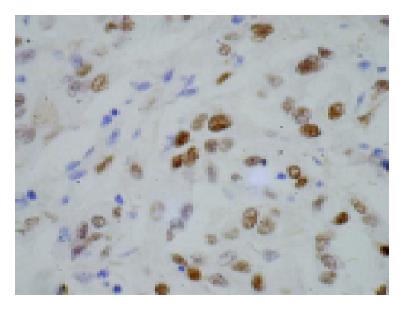
Figure 1 TAp73α protein in HCC, nuclei of tumor cells were stained with yellow or brown (×400).
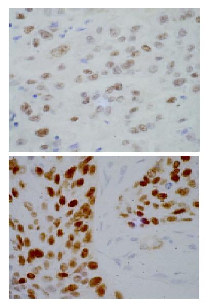
Figure 2 p53 protein in HCC, nuclei of tumor cells were stained with yellow or brown (×400).
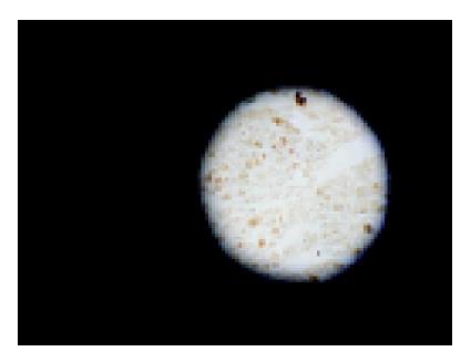
Figure 3 PCNA protein in HCC, nuclei of tumor cells were stained with brown (×400).
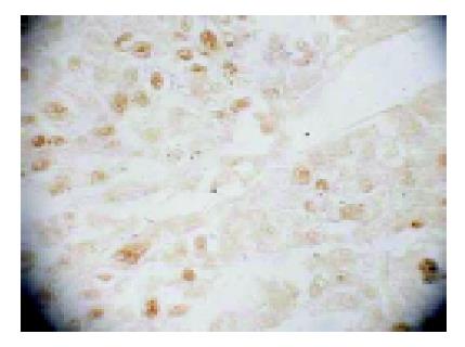
Figure 4 Apoptosis cells were stained in blue, while non-apoptosis cells were stained in pink (×400).
- Citation: Qin HX, Nan KJ, Yang G, Jing Z, Ruan ZP, Li CL, Xu R, Guo H, Sui CG, Wei YC. Expression and clinical significance of TAp73α, p53, PCNA and apoptosis in hepatocellular carcinoma. World J Gastroenterol 2005; 11(18): 2709-2713
- URL: https://www.wjgnet.com/1007-9327/full/v11/i18/2709.htm
- DOI: https://dx.doi.org/10.3748/wjg.v11.i18.2709


