Copyright
©2005 Baishideng Publishing Group Inc.
World J Gastroenterol. May 14, 2005; 11(18): 2697-2703
Published online May 14, 2005. doi: 10.3748/wjg.v11.i18.2697
Published online May 14, 2005. doi: 10.3748/wjg.v11.i18.2697
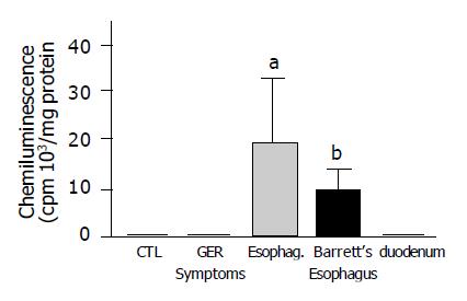
Figure 1 Anion superoxide generation measured by chemiluminescence in human esophageal biopsies with different grades of damage and in normal esophageal and duodenal epithelium.
Chemiluminescence was significantly higher in patients with erosive esophagitis (grade II-IV) and Barrett’s esophagus compared to control patients. (n = 10-28 aP<0.05, bP<0.01 vs normal esophageal mucosa).
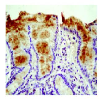
Figure 2 Microphotograph showing nitrotyrosine immunohistochemical staining in a patient with Barrett’s esophagus.
Nitrotyrosine presence was visualized using a specific antibody.
Figure 3 A: SOD activity decreased with damage.
Significant differences were found between patients with esophagitis (grade II-IV) or Barrett’s esophagus and the control group (normal esophageal mucosa) (n = 12-27); B: GSH levels were significantly increased in biopsies from patients with GERD symptoms, esophagitis and duodenal mucosa compared to biopsy samples of normal esophageal mucosa (n = 10-12); C: catalase activity was significantly increased in biopsies from both esophagitis and Barrett’s esophagus patients when compared to normal esophageal mucosa. Duodenal catalase activity was also high (n = 10-12, aP<0.05, bP<0.01 vs normal esophageal mucosa).
Figure 4 Microphotograph of immunohistochemical Cu,ZnSOD staining.
Cu,ZnSOD was present both in control subjects (normal esophageal mucosa) (A) and in patients with esophagitis (B) or Barrett’s esophagus (C), but its expression increased according to the severity of lesion, reaching the highest level in Barrett esophagus.
Figure 5 Representative Western analysis of tissue lysates (22 μg/lane) from control subjects (normal esophageal mucosa) and patients with esophagitis and Barrett’s esophagus.
A: Western blot analysis of Cu,ZnSOD expression in esophageal biopsies demonstrated an increase of Cu,ZnSOD expression in Barrett’s esophagus biopsies (lanes 3-5) compared to control subjects (lanes 1,2) and patients with esophagitis (lanes 6,7); B: A band at 24 ku corresponding to monomeric MnSOD was observed in all groups evaluated. Increased staining for MnSOD was observed in Barrett’s esophagus (lanes 3-5) and esophagitis (lanes 6,7) compared to control patients (lanes 1,2); C: Immunoprecipitated tyrosine-nitrated proteins and subsequent incubation with anti-MnSOD demonstrated that MnSOD nitrated is increased in patients with esophagitis (lanes 6,7) and Barrett’s esophagus (lanes 3-5) compared to normal subjects (lanes 1,2).
- Citation: Jiménez P, Piazuelo E, Sánchez MT, Ortego J, Soteras F, Lanas A. Free radicals and antioxidant systems in reflux esophagitis and Barrett’s esophagus. World J Gastroenterol 2005; 11(18): 2697-2703
- URL: https://www.wjgnet.com/1007-9327/full/v11/i18/2697.htm
- DOI: https://dx.doi.org/10.3748/wjg.v11.i18.2697


