Copyright
©2005 Baishideng Publishing Group Inc.
World J Gastroenterol. May 7, 2005; 11(17): 2687-2689
Published online May 7, 2005. doi: 10.3748/wjg.v11.i17.2687
Published online May 7, 2005. doi: 10.3748/wjg.v11.i17.2687
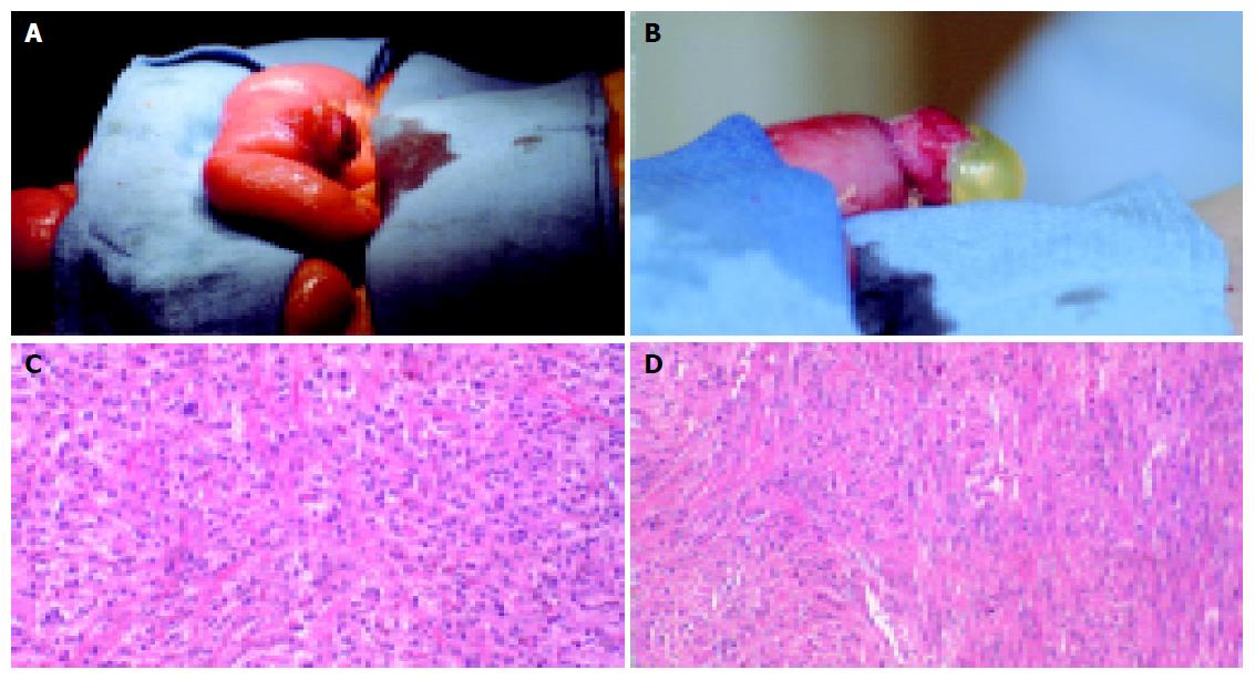
Figure 1 A: Gross photograph of proximal, ruptured tumor located approximately 2 feet distal to the ligament of Treitz.
B: Gross photograph of distal, non-ruptured tumor approximately 2 feet distal to the first lesion. C: Light microscopy (×40) of proximal tumor, revealing malignant neoplasm forming nests and sheets. The tumor extended from the mucosal to the serosal bowel layers. Individual cells showed highly pleomorphic nuclei with prominent, sometimes multiple, nucleoli and abundant pale eosinophilic cytoplasm. D: Light microscopy (×40) of distal tumor showing tumor cells arranged in spindles and arranged in fascicles. There was involvement of the full thickness of the bowel wall. Individual cells showed mildly pleomorphic nuclei and eosinophilic cytoplasm.
- Citation: Brummel N, Awad Z, Frazier S, Liu J, Rangnekar N. Perforation of metastatic melanoma to the small bowel with simultaneous gastrointestinal stromal tumor. World J Gastroenterol 2005; 11(17): 2687-2689
- URL: https://www.wjgnet.com/1007-9327/full/v11/i17/2687.htm
- DOI: https://dx.doi.org/10.3748/wjg.v11.i17.2687


