Copyright
©2005 Baishideng Publishing Group Inc.
World J Gastroenterol. May 7, 2005; 11(17): 2684-2686
Published online May 7, 2005. doi: 10.3748/wjg.v11.i17.2684
Published online May 7, 2005. doi: 10.3748/wjg.v11.i17.2684
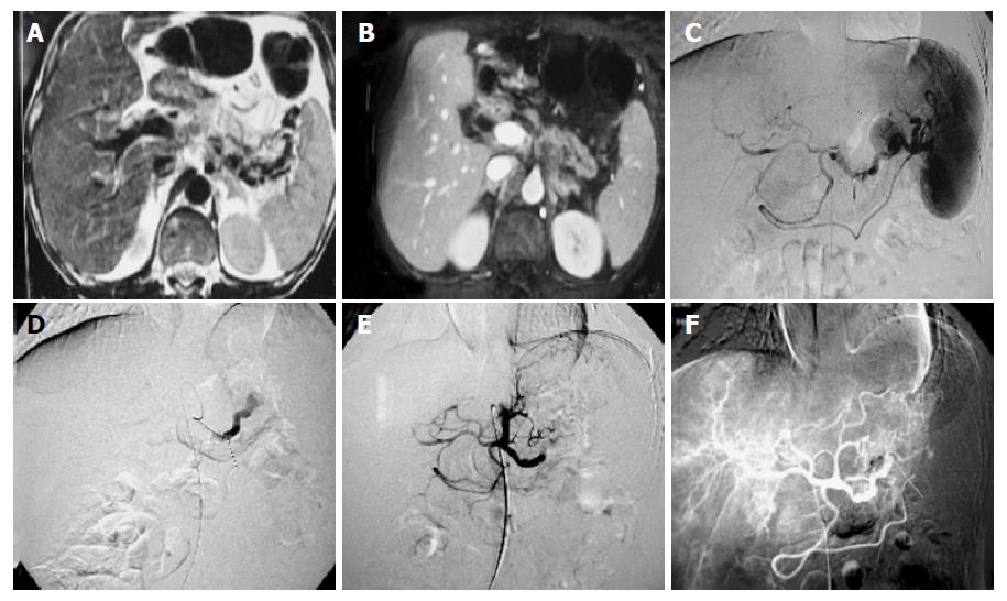
Figure 1 A: Unenhanced abdominal MRI scan 5-mo ago shows varicosis of the hilum of spleen vein and there is no sign of pseudoaneurysm of the splenic artery; B: contrast-enhanced abdominal CT scan shows enhancement of pseudoaneurysm; C: anterior-posterior view of the celiac artery angiogram shows a pseudoaneurysmal sac originating from the splenic artery; D: angiogram shows 3-F SP catheter extending into more distal splenic artery through the 5-F RH catheter; E-F: splenic artery arteriograms after immediatel embolization and 5-d later show the pseudoaneurysmal sac and distal splenic artery stop growing.
- Citation: Guan YS, Sun L, Zhou XP, Li X, Fei ZJ, Zheng XH, He Q. Polyvinyl alcohol and gelatin sponge particle embolization of splenic artery pseudoaneurysm complicating chronic alcoholic pancreatitis. World J Gastroenterol 2005; 11(17): 2684-2686
- URL: https://www.wjgnet.com/1007-9327/full/v11/i17/2684.htm
- DOI: https://dx.doi.org/10.3748/wjg.v11.i17.2684


