Copyright
©2005 Baishideng Publishing Group Inc.
World J Gastroenterol. May 7, 2005; 11(17): 2653-2655
Published online May 7, 2005. doi: 10.3748/wjg.v11.i17.2653
Published online May 7, 2005. doi: 10.3748/wjg.v11.i17.2653
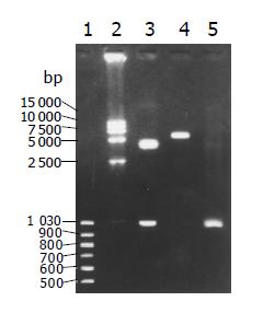
Figure 1 Result of PCR and enzyme pCDNA31hisB-hFasL plasmid lines 1, 2: Molecule marker; line 3: EcoRI and BamHI enzyme cut plasmid; lane 4: Uncut plasmid; lane 5: PCR result The sequence of pCDNA31hisB-hFasLwas tested in GenBank Blast.
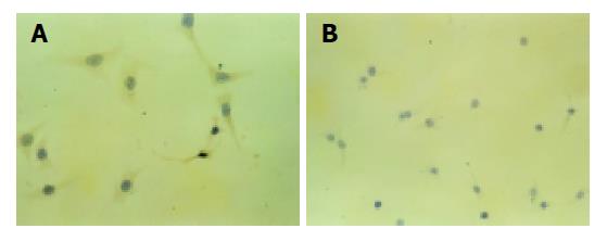
Figure 2 Expression of FasL in HepG2 cells.
A: Transfected FasL cells (×200); B: control cells (×100).
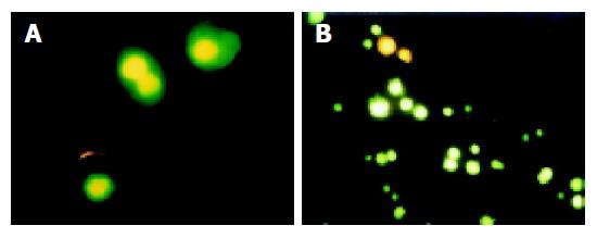
Figure 3 Apoptotic cells at different stages.
A: middle and late stage apoptotic cells; B: early, middle and late stage apoptotic cells.
- Citation: Chen J, Su XS, Jiang YF, Gong GZ, Zheng YH, Li GY. Transfection of apoptosis related gene Fas ligand in human hepatocellular carcinoma cells and its significance in apoptosis. World J Gastroenterol 2005; 11(17): 2653-2655
- URL: https://www.wjgnet.com/1007-9327/full/v11/i17/2653.htm
- DOI: https://dx.doi.org/10.3748/wjg.v11.i17.2653


