Copyright
©2005 Baishideng Publishing Group Inc.
World J Gastroenterol. May 7, 2005; 11(17): 2583-2590
Published online May 7, 2005. doi: 10.3748/wjg.v11.i17.2583
Published online May 7, 2005. doi: 10.3748/wjg.v11.i17.2583
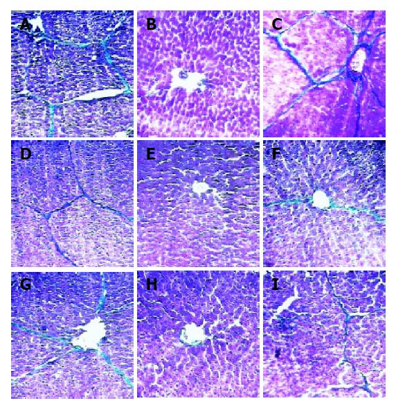
Figure 1 Comparison of liver pathological changes in three groups of pig serum-induced model rats at wk 10 (A-C), 14 (D-F), 20 (G-I) (Masson 100×).
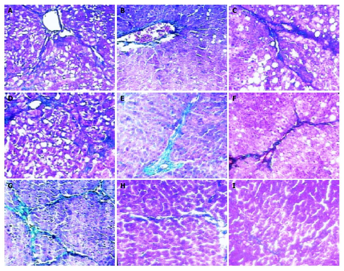
Figure 2 Comparisons of liver pathologic features in three groups of CCl4-induced model rats at wk 10 (A-C), 14 (D-F), 20 (G-I) (Masson, 100×).
Figure 3 Positive rates of type I collagen (A), α-SMA (B) and TIMP-1 mRNA (C) in pig serum-induced model rats at different time points.
Figure 4 Positive rates of type I collagen (A), α-SMA (B) and TIMP-1 mRNA (C) in CCl4 model rats.
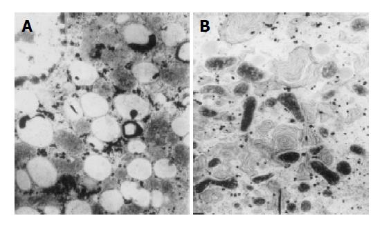
Figure 5 Electron microscopic observation of liver tissues in fibrotic model (A) and prophylaxis model (B).
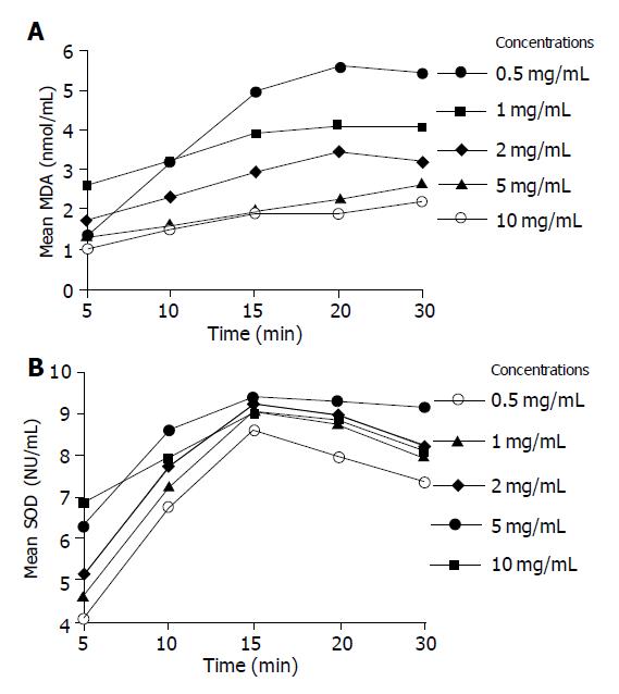
Figure 6 Dose- and time-dependent effect of Radix Salviae Miltiorrhizae on MDA production (A) and SOD activity (B) at different time points and different concentrations of Radix Salviae Miltiorrhizae root extraction.
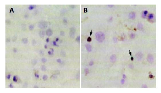
Figure 7 HSCs stained by TUNEL (A) and apoptotic cells characterized by compaction of nuclear chromatin and condensation of cytoplasm (B).
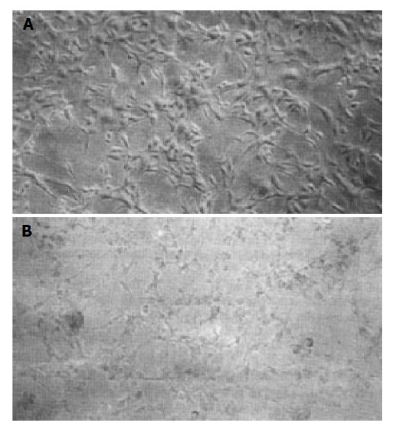
Figure 8 Morphological changes of treated HSCs (A) and untreated HSCs (B) with Yi-gan-kang under light microscope.
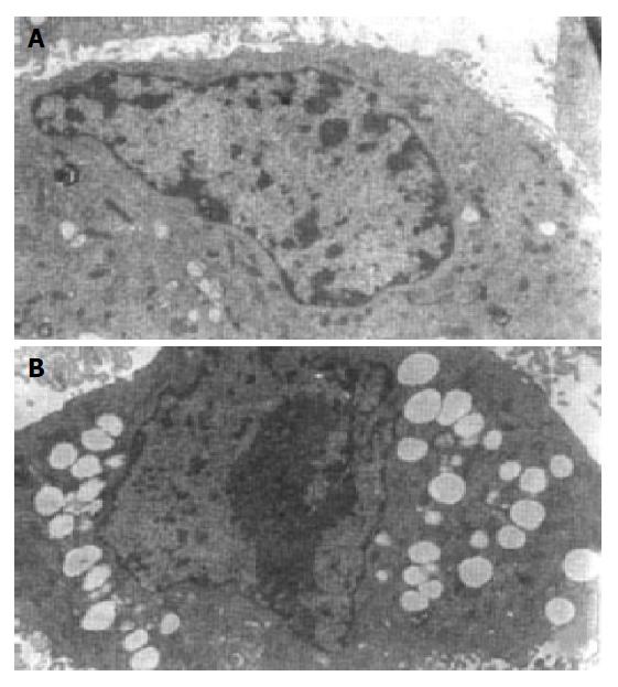
Figure 9 Ultrastructures of treated HSCs (A) and untreated HSCs (B) with Yi-gan-kang.
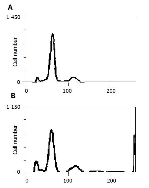
Figure 10 Apoptosis rate of HSCs in control group (A) and Yi-gan-kang decoction treatment group (B).
- Citation: Yao XX, Jiang SL, Tang YW, Yao DM, Yao X. Efficacy of Chinese medicine Yi-gan-kang granule in prophylaxis and treatment of liver fibrosis in rats. World J Gastroenterol 2005; 11(17): 2583-2590
- URL: https://www.wjgnet.com/1007-9327/full/v11/i17/2583.htm
- DOI: https://dx.doi.org/10.3748/wjg.v11.i17.2583


