Copyright
©2005 Baishideng Publishing Group Inc.
World J Gastroenterol. Apr 21, 2005; 11(15): 2367-2369
Published online Apr 21, 2005. doi: 10.3748/wjg.v11.i15.2367
Published online Apr 21, 2005. doi: 10.3748/wjg.v11.i15.2367
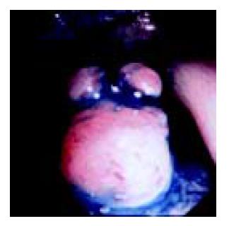
Figure 1 Colonoscopic picture showing a polypoid tumor in the sigmoid colon.
The tumor was covered with normal lining crypts, but had slightly engorged vessels on the surface.
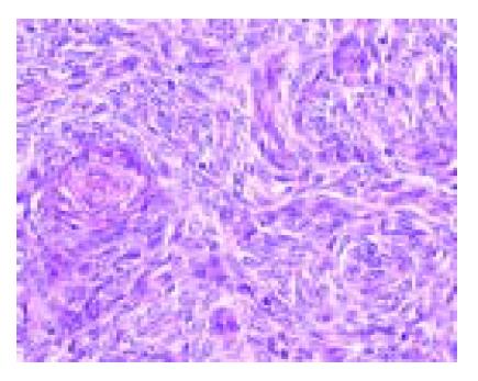
Figure 2 Microscopic picture of the surgical specimen showing dense proliferation of spindle-like cells with ovoid or round nuclei, dispersed chromatin and sparse cytoplasm, arranged in ill-defined whorls centered on small arterioles.
These showed features similar to the primary uterine low-grade ESS.
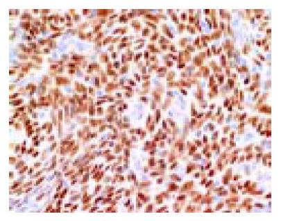
Figure 3 Immunohistochemistry showing strong diffuse immunoreactivity of the nuclei of neoplastic cells for progesterone receptor.
- Citation: Asada Y, Isomoto H, Akama F, Nomura N, Wen CY, Nakao H, Murata I, Toriyama K, Kohno S. Metastatic low-grade endometrial stromal sarcoma of the sigmoid colon three years after hysterectomy. World J Gastroenterol 2005; 11(15): 2367-2369
- URL: https://www.wjgnet.com/1007-9327/full/v11/i15/2367.htm
- DOI: https://dx.doi.org/10.3748/wjg.v11.i15.2367


