Copyright
©2005 Baishideng Publishing Group Inc.
World J Gastroenterol. Apr 21, 2005; 11(15): 2244-2248
Published online Apr 21, 2005. doi: 10.3748/wjg.v11.i15.2244
Published online Apr 21, 2005. doi: 10.3748/wjg.v11.i15.2244
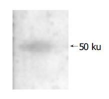
Figure 1 mAb binding to 50 ku ODC protein as shown by Western blotting.
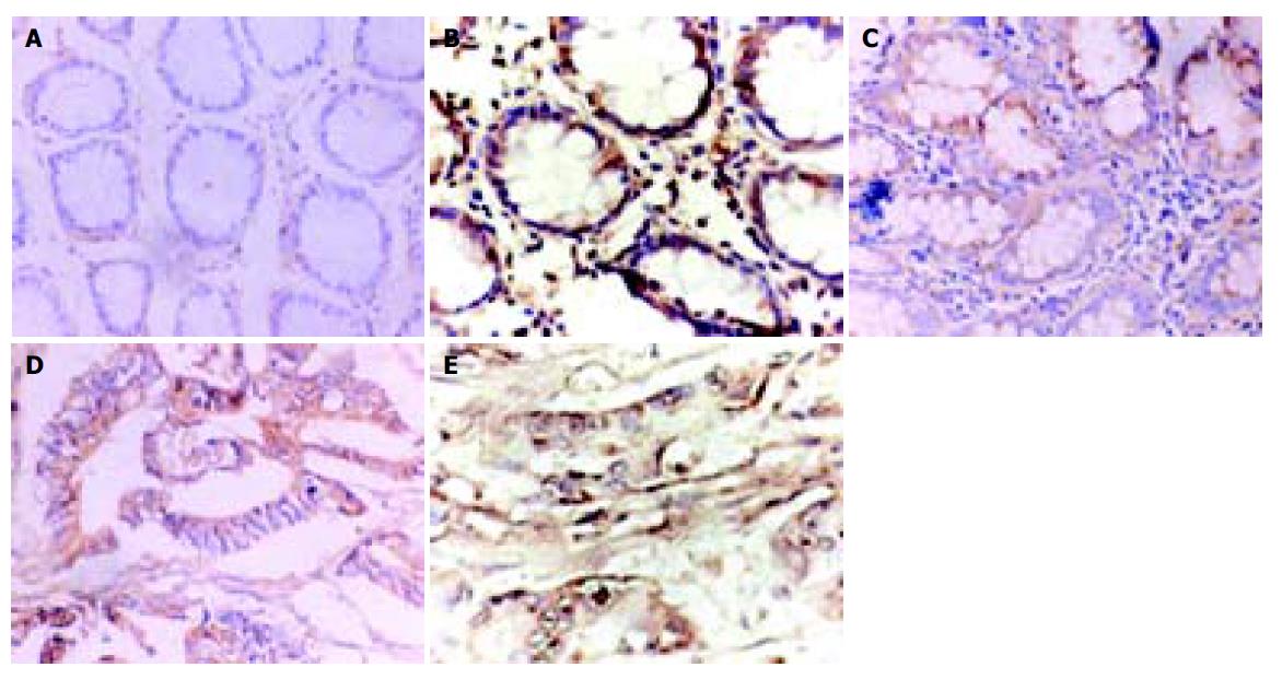
Figure 2 Immunohistochemical staining of colon tissues.
A: Normal colon tissue obtained 15 cm apart from neoplasm; B: Tissue from well-differentiated adenocarcinoma; C: Tissue from moderately differentiated adenocarcinoma; D: Tissue from poorly differentiated mucinous adenocarcinoma; E: Tissue from undifferentiated carcinoma.
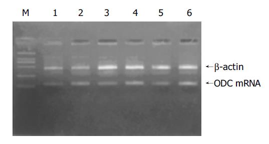
Figure 3 ODC mRNA expression assay by RT-PCR.
M: molecular weight marker DL-2000. Lanes 1, 3 and 5: normal mucosa tissues. Lanes 2, 4 and 6: malignant tissue samples. β-actin was amplified as an internal control.
- Citation: Hu HY, Liu XX, Jiang CY, Lu Y, Liu SL, Bian JF, Wang XM, Geng Z, Zhang Y, Zhang B. Ornithine decarboxylase gene is overexpressed in colorectal carcinoma. World J Gastroenterol 2005; 11(15): 2244-2248
- URL: https://www.wjgnet.com/1007-9327/full/v11/i15/2244.htm
- DOI: https://dx.doi.org/10.3748/wjg.v11.i15.2244


