Copyright
©2005 Baishideng Publishing Group Inc.
World J Gastroenterol. Apr 14, 2005; 11(14): 2203-2205
Published online Apr 14, 2005. doi: 10.3748/wjg.v11.i14.2203
Published online Apr 14, 2005. doi: 10.3748/wjg.v11.i14.2203
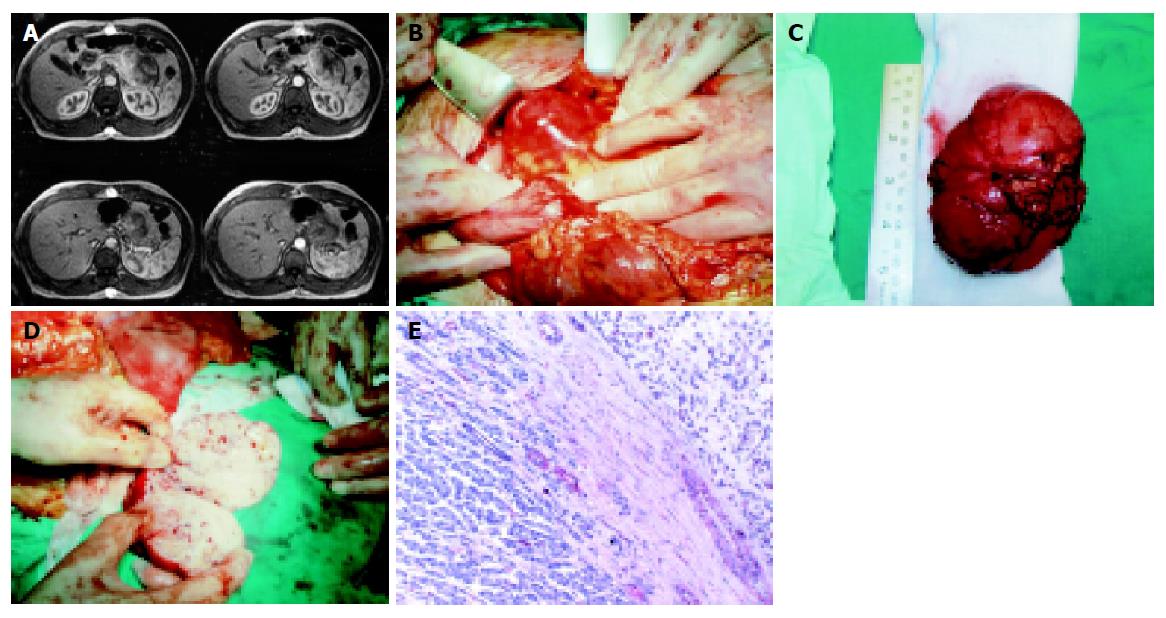
Figure 1 A: The abdominal CT scan revealing the pancreatic metastatic lesion; B: The pancreatic metastatic lesion while excised; C: The excised surgical specimen; D: A section of the surgical specimen; E: Normal pancreatic tissue (1) and tumor cells of the metastatic EMC (2) separated by connective tissue (× 400).
- Citation: Fotiadis C, Charalambopoulos A, Chatzikokolis S, Zografos G, Genetzakis M, Tringidou R. Extraskeletal myxoid chondrosarcoma metastatic to the pancreas: A case report. World J Gastroenterol 2005; 11(14): 2203-2205
- URL: https://www.wjgnet.com/1007-9327/full/v11/i14/2203.htm
- DOI: https://dx.doi.org/10.3748/wjg.v11.i14.2203


