Copyright
©2005 Baishideng Publishing Group Inc.
World J Gastroenterol. Apr 14, 2005; 11(14): 2157-2161
Published online Apr 14, 2005. doi: 10.3748/wjg.v11.i14.2157
Published online Apr 14, 2005. doi: 10.3748/wjg.v11.i14.2157
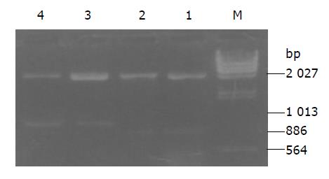
Figure 1 Agarose gel electrophoresis analysis of pRcHCN3-3’ and pRcHCN3-5’ digested with XbaI.
Lane M: DNA marker (λDNA/Hind III). Lanes 1 and 2: pRcHCNS3-5’ plasmid, 886-bp fragment. Lanes 3 and 4: pRcHCNS3-3’ plasmid, 1 031-bp fragment.
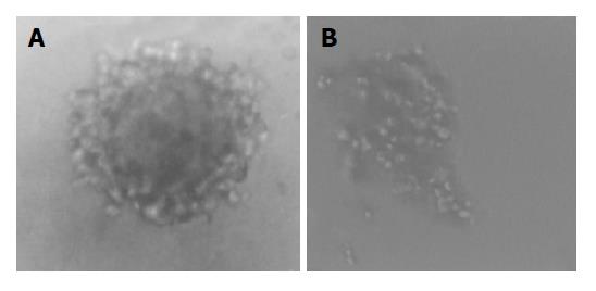
Figure 2 Clones of QSG7701 cell transfected with pRcHCNS3-5’ and pRcHCN3-3’ plasmid in soft agar.
A: pRcHCNS3-5’ transfected group. B: pRcHCNS3-3’ transfected group (×100).
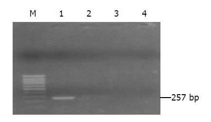
Figure 3 Identification of expression plasmid of HCNS3 protein in QSG7701 cells.
Lane 1: pRcHCNS3-5’; lane 2: pRcCMV; lane 3: untransfected QSG7701 cells; lane 4: blank control.
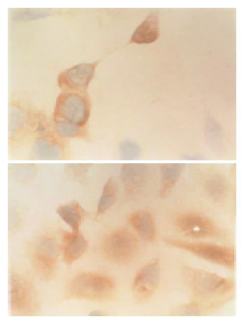
Figure 4 Expression of HCV NS3 protein in QSG77 cell transfected with pRcHCNS3-5’ plasmid.
The positive products were localized in cytoplasm. SP ×400.
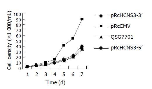
Figure 5 Growth curve of four kinds of cells.
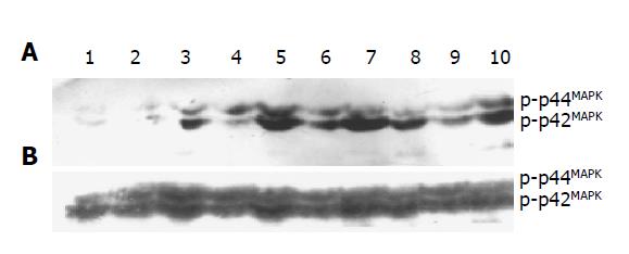
Figure 6 Western blot analysis of phosphorylated (A) and non-phosphorylated (B) MAPK in each group.
Lanes 1 and 2: non-transfected group; Lanes 3 and 4: pRcCMV transfected group; Lanes 5-7: pRcHCNS3-5’ transfected group; Lanes 8-10: pRcHCNS3-3’ transfected group.
- Citation: Feng DY, Sun Y, Cheng RX, Ouyang XM, Zheng H. Effect of hepatitis C virus nonstructural protein NS3 on proliferation and MAPK phosphorylation of normal hepatocyte line. World J Gastroenterol 2005; 11(14): 2157-2161
- URL: https://www.wjgnet.com/1007-9327/full/v11/i14/2157.htm
- DOI: https://dx.doi.org/10.3748/wjg.v11.i14.2157


