Copyright
©2005 Baishideng Publishing Group Inc.
World J Gastroenterol. Apr 14, 2005; 11(14): 2136-2141
Published online Apr 14, 2005. doi: 10.3748/wjg.v11.i14.2136
Published online Apr 14, 2005. doi: 10.3748/wjg.v11.i14.2136
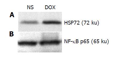
Figure 1 Western blot analysis of HSP72 (A) and NF-κB p65 subunit (B) in liver cytoplasmic extracts before SFC in NS and DOX groups.
Figure 2 Western blot analysis of NF-κB presentation (A) in liver nuclear extracts, IκB-α expression (B) and immunoprecipitation/Western blot analysis of p-IκB-α expression (C) in liver cytoplasmic extracts at different time points, i.
e. before SFC, at the end of SFC (20 min), 30 min after the restoration of the abdominal circulation, 3 h after the restoration of the abdominal circulation, 6 h after the restoration of the abdominal circulation. WB, Western blot, IP, Immunoprecipitation.
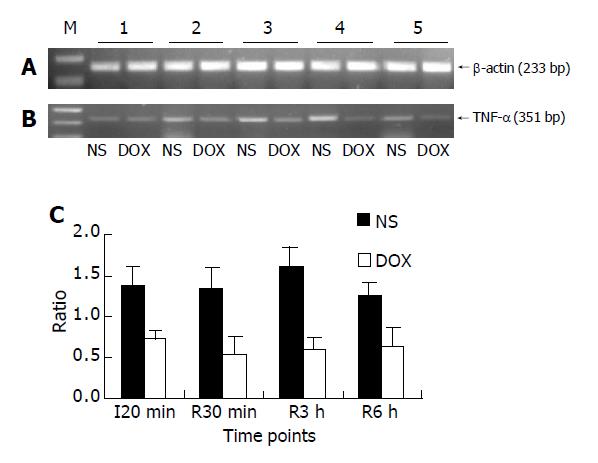
Figure 3 Semiquantitive RT-PCR analysis of β-actin (A) as an internal control and TNF-α mRNA (B) expression in liver tissues at different time points, i.
e., before SFC, at the end of SFC (20 min), 30 min after the restoration of the abdominal circulation, 3 h after the restoration of the abdominal circulation, 6 h after the restoration of the abdominal circulation, and the ratio between the expression of TNF-α mRNA at different time points and the expression of TNF-α mRNA (C).
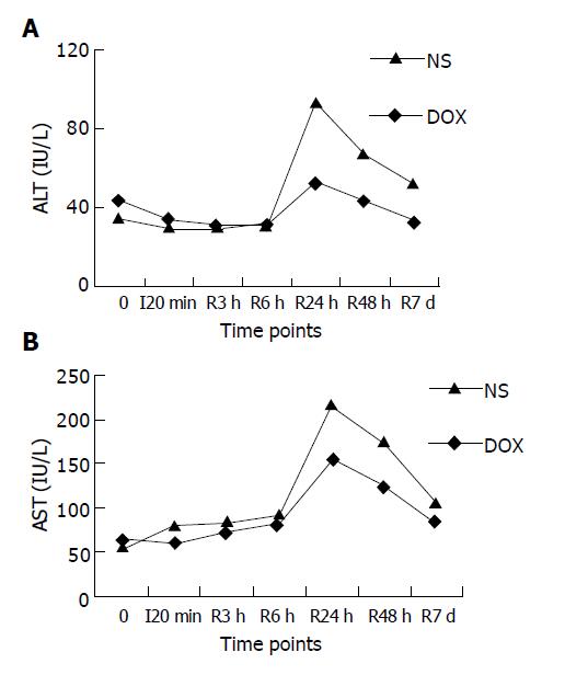
Figure 4 Serum level of ALT (A) and AST (B) at different time points before and after SFC respectively, 0, before SFC, I20 min, at the end of SFC (20 min), R3h, 3 h after the restoration of the abdominal circulation, R6 h, 6 h after the restoration of the abdominal circulation, R24 h, 24 h after the restoration of the abdominal circulation, R48 h, 48 h after the restoration of the abdominal circulation, R7d, 7 d after the restoration of the abdominal circulation.
- Citation: Lu H, Zhu ZG, Yao XX, Zhao R, Yan C, Zhang Y, Liu BY, Yin HR, Lin YZ. Hepatic preconditioning of doxorubicin in stop-flow chemotherapy: NF-κB/IκB-α pathway and expression of HSP72. World J Gastroenterol 2005; 11(14): 2136-2141
- URL: https://www.wjgnet.com/1007-9327/full/v11/i14/2136.htm
- DOI: https://dx.doi.org/10.3748/wjg.v11.i14.2136


