Copyright
©2005 Baishideng Publishing Group Inc.
World J Gastroenterol. Apr 14, 2005; 11(14): 2055-2060
Published online Apr 14, 2005. doi: 10.3748/wjg.v11.i14.2055
Published online Apr 14, 2005. doi: 10.3748/wjg.v11.i14.2055
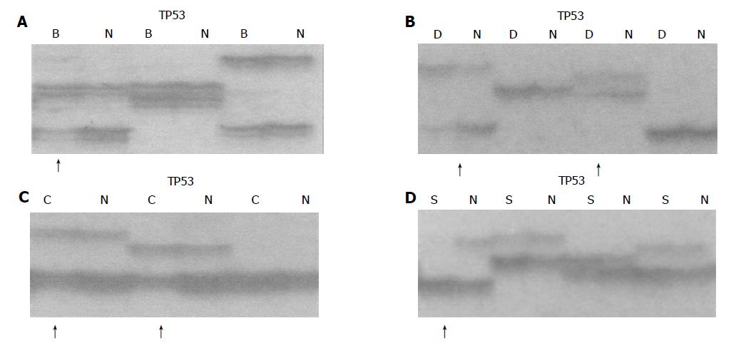
Figure 1 Allelic deletion patterns in TP53 microsatellite loci.
Arrows indicate LOH in different lesions. B: basal cell hyperplasia (BCH), D: dysplasia (DYS), C: carcinoma in situ (CIS), S: squamous cell carcinoma(SCC), N: normal tissue, A: LOH in BCH; B: LOH in DYS; C: LOH in CIS; D: LOH in SCC.
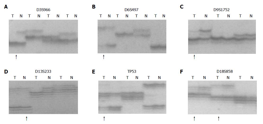
Figure 2 Allelic deletion patterns of various microsatellite markers in ESCC.
Paired tumor DNA (T) and non-neoplastic DNA (N) were examined for each case. Arrows indicate LOH in tumor samples. A: D3S966 LOH; B: D6S497 LOH; C: D9S1752 LOH; D: D13S233 LOH; E: TP53 LOH; F: D18S858 LOH.
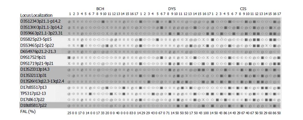
Figure 3 Distributions of microsatellite DNA-LOH in different precancerous lesions of esophagus FAL: fractional allelic loss for each tumor ○ retention of heterozygosity ■ loss of heterozygosity Ø uninformative, X not done.
- Citation: An JY, Fan ZM, Gao SS, Zhuang ZH, Qin YR, Li JL, He X, Tsao GSW, Wang LD. Loss of heterozygosity in multistage carcinogenesis of esophageal carcinoma at high-incidence area in Henan Province, China. World J Gastroenterol 2005; 11(14): 2055-2060
- URL: https://www.wjgnet.com/1007-9327/full/v11/i14/2055.htm
- DOI: https://dx.doi.org/10.3748/wjg.v11.i14.2055


