Copyright
©2005 Baishideng Publishing Group Inc.
World J Gastroenterol. Mar 28, 2005; 11(12): 1886-1889
Published online Mar 28, 2005. doi: 10.3748/wjg.v11.i12.1886
Published online Mar 28, 2005. doi: 10.3748/wjg.v11.i12.1886
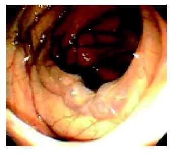
Figure 1 This endoscopic image of the colon revealed varices (arrows) over the hepatic flexure.
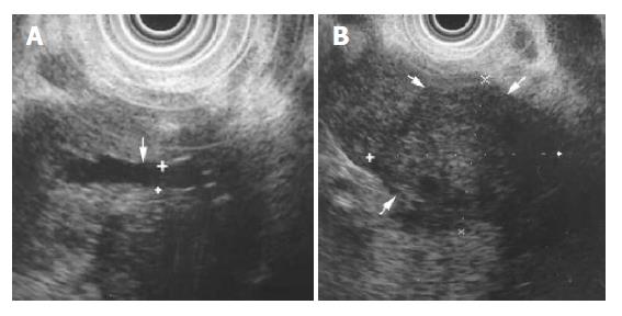
Figure 2 EUS revealed mild dilation of the main pancreatic duct (arrow in A) and a solid mass over pancreatic head, measuring approximately 3.
4 cm by 2.8 cm (arrows in B).
Figure 3 Abdominal computed tomography of the arterial phase revealed patent splenic vein (arrow in A).
With the disappearance of intravenous contrast in the superior mesenteric vein (black arrow in B), but patent superior mesenteric artery (white arrow in B), and the mass (white cross in B) over the uncinate pancreatic head area becomes apparent. Prominent collateral circulation (arrow in C) could be found in abdominal cavity near the hepatic flexure.
- Citation: Ho YP, Lin CJ, Su MY, Tseng JH, Chiu CT, Chen PC. Isolated varices over hepatic flexure colon indicating superior mesenteric venous thrombosis caused by uncinate pancreatic head cancer - a case report. World J Gastroenterol 2005; 11(12): 1886-1889
- URL: https://www.wjgnet.com/1007-9327/full/v11/i12/1886.htm
- DOI: https://dx.doi.org/10.3748/wjg.v11.i12.1886


