Copyright
©2005 Baishideng Publishing Group Inc.
World J Gastroenterol. Mar 28, 2005; 11(12): 1881-1883
Published online Mar 28, 2005. doi: 10.3748/wjg.v11.i12.1881
Published online Mar 28, 2005. doi: 10.3748/wjg.v11.i12.1881
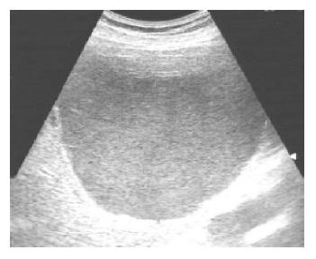
Figure 1 Homogenous hyper-echoic material filling a solitary liver cyst.
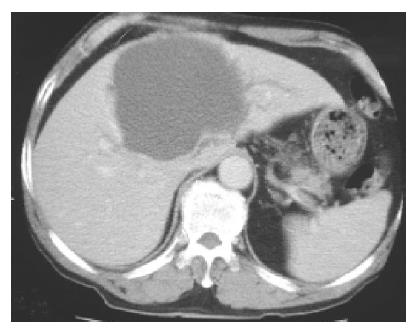
Figure 2 Contrast-enhanced CT showing a huge liver cyst causing mild intra-hepatic duct dilatation.
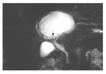
Figure 3 A T2-weighted MR image reveals a mass consisting mostly of fluid density.
The lower portion has low signal intensity (arrow), suggesting cyst hemorrhage or a cystic tumor.
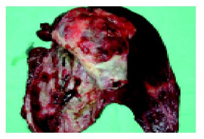
Figure 4 Opened cystic tumor which showed several dark red slight papillary growths and small dome-shaped nodules (arrow) on the inner surface.
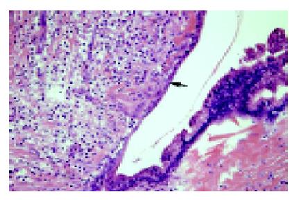
Figure 5 The cystic wall is lined by a single layer of biliary epithelium (arrow) with focal proliferation, a slight papillary appearance, and mild nuclear atypia.
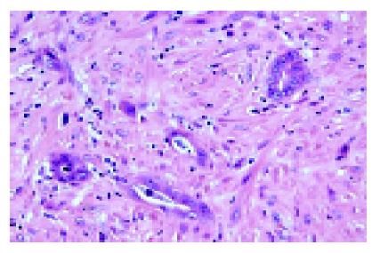
Figure 6 Histology of a nodule.
Small round or angulated glandular structures, small cords, and plump individual epithelial cells are noted. The cells have nuclear atypia, hyperchromatism, and enlargement.
- Citation: Lin CC, Lin SC, Ko WC, Chang KM, Shih SC. Adenocarcinoma and infection in a solitary hepatic cyst: A case report. World J Gastroenterol 2005; 11(12): 1881-1883
- URL: https://www.wjgnet.com/1007-9327/full/v11/i12/1881.htm
- DOI: https://dx.doi.org/10.3748/wjg.v11.i12.1881


