Copyright
©2005 Baishideng Publishing Group Inc.
World J Gastroenterol. Mar 21, 2005; 11(11): 1709-1711
Published online Mar 21, 2005. doi: 10.3748/wjg.v11.i11.1709
Published online Mar 21, 2005. doi: 10.3748/wjg.v11.i11.1709
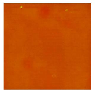
Figure 1 Some irregular granules were scanned in 5% dextrose.
Scan size was 50 μm.
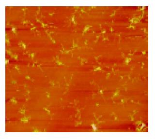
Figure 2 Finely branched granules were observed in PBS buffer.
Scan size was 50 μm.
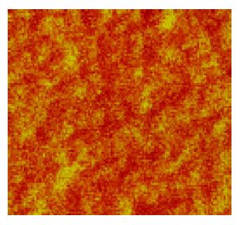
Figure 3 Fine structures of human erythrocyte membranes featured with the shape of pores and protrusions.
The erythrocyte was mixed with 5% dextrose. Scan size was 500 nm.
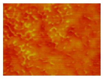
Figure 4 Fine structures of human erythrocyte membranes is shown.
The erythrocyte was mixed with PBS buffer. A lot of granules existed in the image. Scan size was 500 nm.
- Citation: Ji XL, Ma YM, Yin T, Shen MS, Xu X, Guan W. Application of atomic force microscopy in blood research. World J Gastroenterol 2005; 11(11): 1709-1711
- URL: https://www.wjgnet.com/1007-9327/full/v11/i11/1709.htm
- DOI: https://dx.doi.org/10.3748/wjg.v11.i11.1709


