Copyright
©2005 Baishideng Publishing Group Inc.
World J Gastroenterol. Mar 14, 2005; 11(10): 1532-1534
Published online Mar 14, 2005. doi: 10.3748/wjg.v11.i10.1532
Published online Mar 14, 2005. doi: 10.3748/wjg.v11.i10.1532
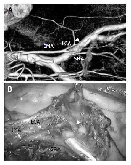
Figure 1 A case of sigmoid colon cancer who underwent a laparoscopic sigmoidectomy.
A: Three-dimensional volume-rendered CT (3DCT) angiographic reconstructions show the IMA and its branches; B: The intraoperative findings similar to the preoperative visualization of the IMA were obtained. White arrowheads show the sigmoidal artery.
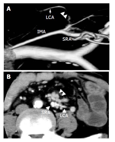
Figure 2 A: 3DCT image of the LCA is partially indistinct (two arrowheads); B: Swollen lymph nodes adhered to the LCA (two arrowheads).
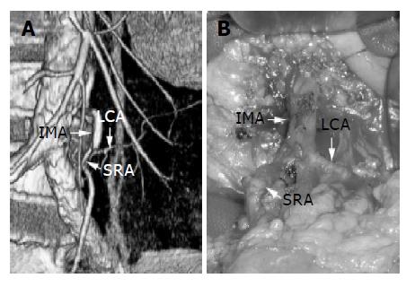
Figure 3 A: A slight deformity of the SRA was showed by 3DCT; B: In intraoperative findings, a slight deformity of the SRA was comfirmed.
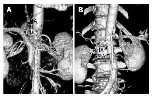
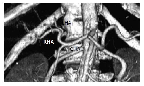
Figure 4 3DCT scans showed the CHA diverging from the SMA (A) and the RHA branching from the SMA (B).
Figure 5 A case of hepatocellular carcinoma who could not undergo angiography due to severe arteriosclerosis.
Preoperative visualization of the HA was obtained.
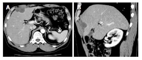
Figure 6 The follow-up patient who underwent right hemicolectomy for ascending colon cancer: A: In the axial view, it was difficult to distinguish whether low density area (LDA) in the lateral segment of the liver was dissemination or liver metastasis; B: The LDA was diagnosed as dissemination by MPR image (a sagittal view).
- Citation: Ohtani H, Kawajiri H, Arimoto Y, Ohno K, Fujimoto Y, Oba H, Adachi K, Hirano M, Terakawa S, Tsubakimoto M. Efficacy of multislice computed tomography for gastroenteric and hepatic surgeries. World J Gastroenterol 2005; 11(10): 1532-1534
- URL: https://www.wjgnet.com/1007-9327/full/v11/i10/1532.htm
- DOI: https://dx.doi.org/10.3748/wjg.v11.i10.1532


