Copyright
©2005 Baishideng Publishing Group Inc.
World J Gastroenterol. Mar 14, 2005; 11(10): 1521-1528
Published online Mar 14, 2005. doi: 10.3748/wjg.v11.i10.1521
Published online Mar 14, 2005. doi: 10.3748/wjg.v11.i10.1521
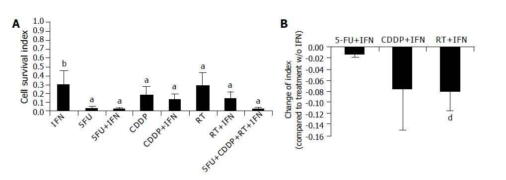
Figure 1 Influence on cell survival Equal amount of cells were seeded out on day +1.
A: Numbers of viable cells were determined after five-day treatment. Cell numbers of untreated cells were set to ‘1’ and cell survival index was calculated. B: Shows the influence of IFN-α in combination treatments. Values of treatments without IFN-α were subtracted from values after combination treatment. Data is shown as mean±SD error from eight cell lines with at least three separate experiments. aP<0.05 was labeled with an asterisk; bP<0.01 with two asterisks and dP<0.001 with three asterisks.
Figure 2 Induction of apoptosis Apoptosis was determined after five-day treatment by staining for AnnexinV+ using flow cytometry (A).
Late apoptosis is defined as AnnexinV+/PI+, necrosis as PI+. B) shows the influence of IFN-α in combination treatments. Values of treatments without IFN-α were subtracted from values after combination treatment. Data is shown as mean±SD error from eight cell lines with at least three separate experiments. aP<0.05 was labeled with an asterisk; bP<0.01 with two asterisks and dP<0.001 with three asterisks.
Figure 3 Inhibition of proliferation Proliferation rate was determined after five-day treatment by using a non-radioactive MTT assay.
Proliferation rate of untreated cells was set to ‘1’ and percentage of proliferation was calculated (A). B) shows the influence of IFN-α in combination treatments. Values of treatments without IFN-α were subtracted from values after combination treatment. Data is shown as mean±standard error from eight cell lines with at least three separate experiments. aP<0.05 was labeled with an asterisk; bP<0.01 with two asterisks and dP<0.001 with three asterisks.
Figure 4 Secretion of VEGF Cells were seeded out after four-day treatment in a density of 1×10⁵ living cells in 2 mL.
Supernatants from treated and untreated tumor cells were collected after 24 h and stored at -80 °C. VEGF concentration was determined by ELISA (A). B) shows the influence of IFN-α in combination treatments. Values of treatments without IFN-α were subtracted from values after combination treatment. Data from three cell lines are shown as mean±standard error from at least three separate experiments. aP<0.05 was labeled with an asterisk and bP<0.01 with two asterisks.
Figure 5 Immunophenotyping of tumor cells Tumor cells were treated as described.
Expression of MHC molecules was analyzed after five-day treatment by flow cytometry (A). B) shows the influence of IFN-α in combination treatments. Values of treatments without IFN-α were subtracted from values after combination treatment. Data is shown as mean±SD error from eight cell lines each with at least four separate experiments. aP<0.05 was labeled with an asterisk and bP<0.01 with two asterisks.
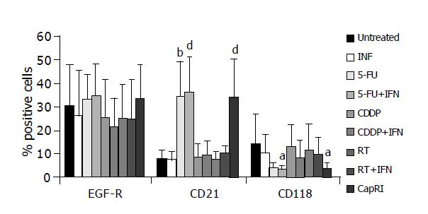
Figure 6 Expression of surface receptors Tumor cells were treated as described.
Expressions of IFN and EGF receptors were analyzed after five-day treatment by flow cytometry. Data is shown as mean±SD error from eight cell lines each with at least four separate experiments. aP<0.05 was labeled with an asterisk; bP<0.01 with two asterisks and dP<0.001 with three asterisks.
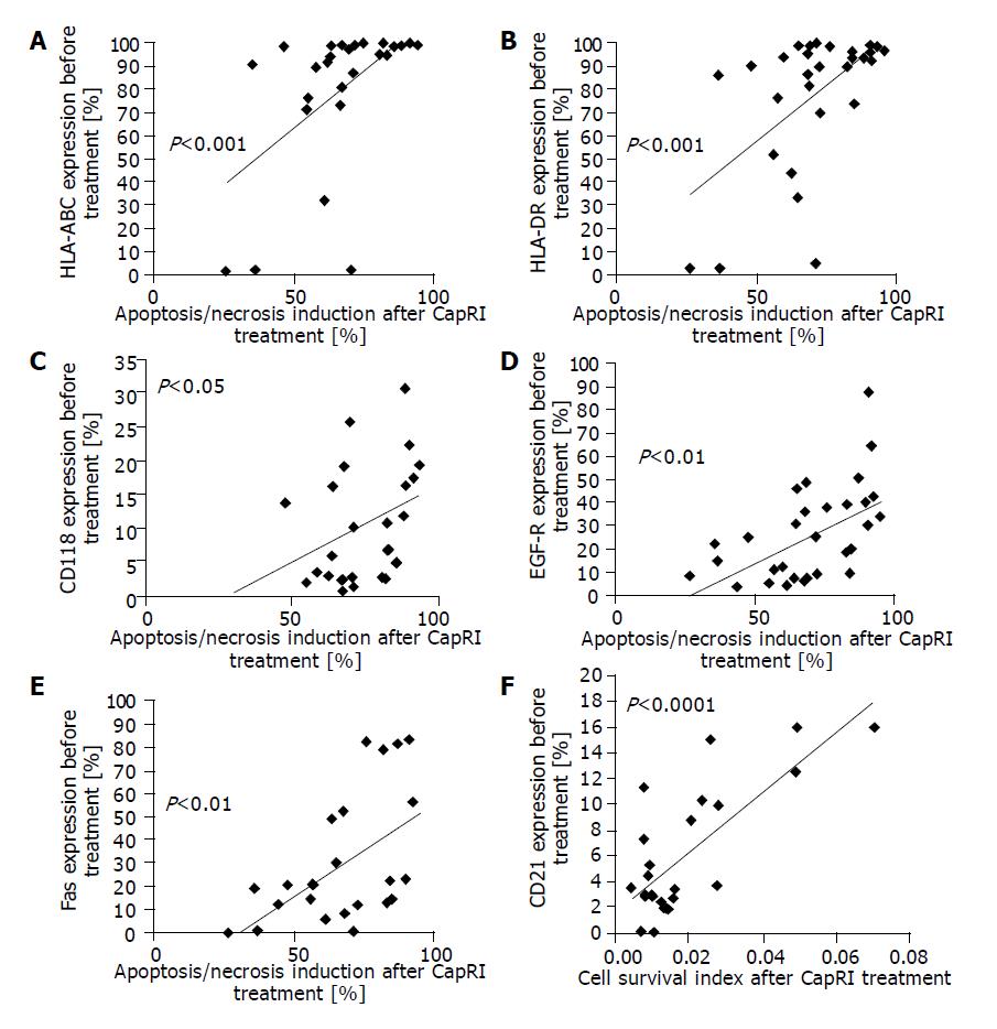
Figure 7 Statistical analysis Expression level on untreated cells were correlated with results from AnnexinV/PI stain, proliferation and cell survival index after treatment with the CapRI scheme and analyzed for use as predictive marker (A-F).
- Citation: Ma JH, Patrut E, Schmidt J, Knaebel HP, Büchler MW, Märten A. Synergistic effects of interferon-alpha in combination with chemoradiation on human pancreatic adenocarcinoma. World J Gastroenterol 2005; 11(10): 1521-1528
- URL: https://www.wjgnet.com/1007-9327/full/v11/i10/1521.htm
- DOI: https://dx.doi.org/10.3748/wjg.v11.i10.1521


