Copyright
©The Author(s) 2004.
World J Gastroenterol. Apr 15, 2004; 10(8): 1191-1197
Published online Apr 15, 2004. doi: 10.3748/wjg.v10.i8.1191
Published online Apr 15, 2004. doi: 10.3748/wjg.v10.i8.1191
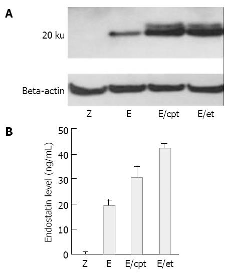
Figure 1 Effects of topoisomerase inhibitors on rAAV mediated endostatin expression level.
Hepa1c1c7 mouse hepatoma cells were transduced with 1 × 10⁴ particles/cell of rAAV-endostatin or rAAV-LacZ. In pretreatment group, etoposide (3 µmol/L) or camptothecin (10 µmol/L) was administered 6 h before transduction. Fourty-eight hours later, the expression of endostatin was determined. A: Analysis of protein expression by NuPAGE electrophoresis, B: Concentration of endostatin measured by ELISA (P < 0.05, rAAV-endostatin in combination with pretreatment groups versus other groups). Z: rAAV-LacZ without pretreatment, E: rAAV-endostatin without pretreatment, E/et: rAAV-endostatin pretreated with etoposide, E/cpt: rAAV-endostatin pretreated with camptothecin.
Figure 2 In vitro biological activities of expressed endostatin.
A: 5 × 10³ HUVECs in a 96-well were cultured in the conditioned media from Hepa1c1c7 mouse hepatoma cells without pre-treatment (Z), those from the cells pretreated with etoposide (Z/et) or camptothecin (Z/cpt), those from the rAAV-endostatin transduced cells without pretreatment (E), those from rAAV-endostatin transduced cells pretreated with etoposide (E/et) or camptothecin (E/cpt). The number of cells was then calculated by a MTT assay. Each value represents mean ± SD of 3 independent experiments (P < 0.05, rAAV-endostatin in combination with pretreatment groups versus other groups). B: Impact on tube formation of endothelial cells. HUVECs were seeded into 24-well plates coated with Matrigel at a density of 5 × 10⁴ cells in each well and cultured in the conditioned media. After 18 h incubation, the level of cell growth and differen-tiation was observed. All tests were performed in triplicate.
Figure 3 Effect of rAAV-endostatin in combination with etoposide on murine sarcoma bearing mice.
Twenty five mice bearing S-180 murine sarcoma cells were randomly divided into 5 groups, namely no treatment, rAAV-LacZ alone, rAAV-LaZ plus etoposide pretreatment, rAAV-endostatin alone, and rAAV-endostatin plus etoposide pretreatment. In the pretreat-ment group, etoposide (40 mg/kg) was administered 3 times for one week by an intraperitoneal injection beginning 7 d prior to rAAV injection, and 1.5 × 10¹² viral particles of rAAV- rAAVendostatin vector were injected into the spleen simultaneously with tumor cell inoculation (5 × 10⁶ S-180 cells) into the liver. The tumor volume was determined 7 d after injecting murine sarcoma cells (P < 0.05, rAAV plus etoposide group versus other groups). Tumor volume = (Length×width2)/2.
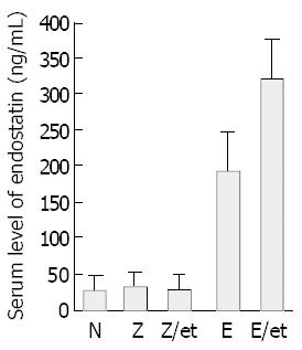
Figure 4 Mouse endostatin levels determined in sera of mice inoculated with murine sarcoma cells.
S-180 murine sarcoma cells were inoculated into liver. Seven days later, ELISA determined the endostatin concentration and the results were expressed as mean ± SD of 5 animals (P < 0.05, rAAV-endostatin plus etoposide group versus other groups).
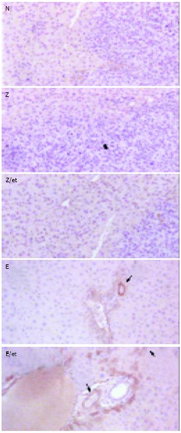
Figure 5 Detection of endostatin in livers of tumor-bearing mice.
Hepatic tumors were induced by injecting murine sar-coma cells into the liver. After 7 d, the livers were harvested from the mice of different groups and analyzed by immuno-histochemical staining for endostatin. Livers without treatment, rAAV-LacZ alone and rAAV-LacZ plus etoposide pretreatment did not express endostatin. The vessels of livers treated with rAAV-endostatin were stained with anti-endostatin antibodies and a significant increase was observed in the rAAV-endostatin plus etoposide treatment group (arrow). Endostatin was also detected in hepatocytes of the rAAV-endostatin plus etoposide treatment group (arrow head).
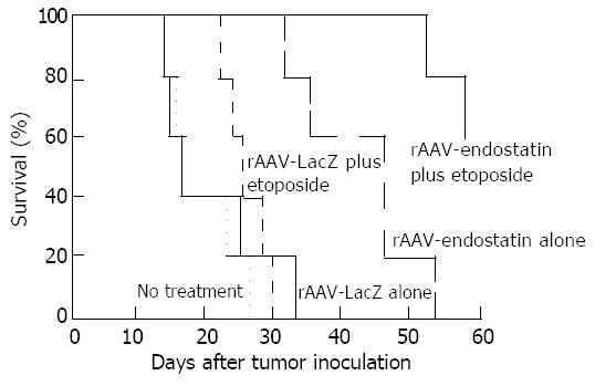
Figure 6 Survival time of sarcoma-bearing mice.
Twenty-five mice were randomly divided into 5 groups: no treatment, rAAV-LacZ alone, rAAV-LacZ plus etoposide, rAAV-endostatin alone, or rAAV-endostatin plus etoposide. The tu-mor-bearing mice treated with rAAV-endostatin in combina-tion with etoposide had a significantly longer survival than those in other groups (P < 0.05).
- Citation: Hong SY, Lee MH, Kim KS, Jung HC, Roh JK, Hyung WJ, Noh SH, Choi SH. Adeno-associated virus mediated endostatin gene therapy in combination with topoisomerase inhibitor effectively controls liver tumor in mouse model. World J Gastroenterol 2004; 10(8): 1191-1197
- URL: https://www.wjgnet.com/1007-9327/full/v10/i8/1191.htm
- DOI: https://dx.doi.org/10.3748/wjg.v10.i8.1191


