Copyright
©The Author(s) 2004.
World J Gastroenterol. Oct 15, 2004; 10(20): 3076-3077
Published online Oct 15, 2004. doi: 10.3748/wjg.v10.i20.3076
Published online Oct 15, 2004. doi: 10.3748/wjg.v10.i20.3076
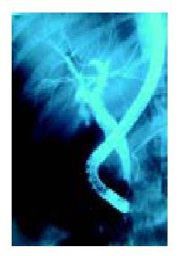
Figure 1 Multiple oval shaped filling defects on cholangiogram.
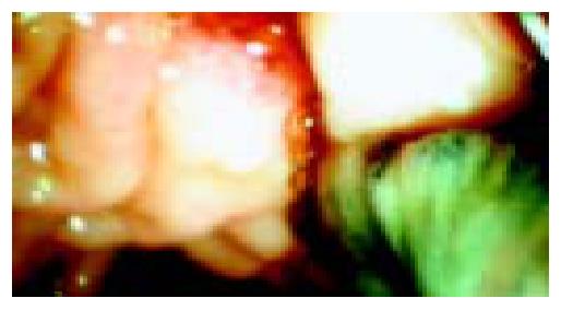
Figure 2 Fasciola hepatica next to papilla of Vater after sphinc-terotomy and balloon extraction.
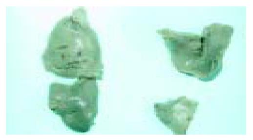
Figure 3 Adult flukes measuring 2.
5 cm × 1.5 cm in diameter.
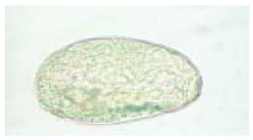
Figure 4 Ova of Fasciola hepatica ( × 40).
- Citation: Dobrucali A, Yigitbasi R, Erzin Y, Sunamak O, Polat E, Yakar H. Fasciola hepatica infestation as a very rare cause of extrahepatic cholestasis. World J Gastroenterol 2004; 10(20): 3076-3077
- URL: https://www.wjgnet.com/1007-9327/full/v10/i20/3076.htm
- DOI: https://dx.doi.org/10.3748/wjg.v10.i20.3076


