Copyright
©The Author(s) 2004.
World J Gastroenterol. Oct 15, 2004; 10(20): 2958-2962
Published online Oct 15, 2004. doi: 10.3748/wjg.v10.i20.2958
Published online Oct 15, 2004. doi: 10.3748/wjg.v10.i20.2958
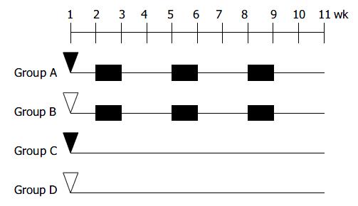
Figure 1 Experimental protocol for inducing colitis-related colon cancer in mice.
▼DMH, 20 mg/kg body mass, intraperi-toneal injection;▽ Saline 0.5 mL/mice intraperitoneal injecton; 30 g/L ■DSS in drinking water.
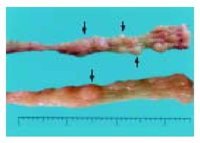
Figure 2 Induced colorectal tumors.
Gross polypoid lesions are evident on the distal and middle portions of the large intestine (arrows).
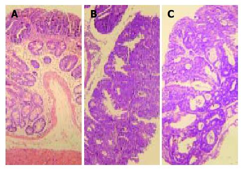
Figure 3 Histopathology showing dysplastic and cancer le-sions developed in the colon of mice from group A.
A: Whole-mount view of DALM with low-grade dysplasia, B: A slightly or non-elevated lesion diagnosed as high-grade dysplasia, C: whole-mount view of an early invasive cancer arising out of DALM. Hematoxylin and eosin stain. Original magnification, × 10.
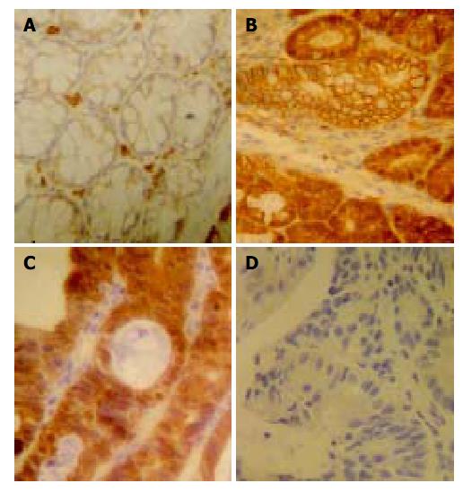
Figure 4 Immunohistochemistry of β -catenin and p53 in dys-plastic lesions found in the colon of mice from group A.
A: Control colon. β -catenin was expressed exclusively on the cell membrane. No cytoplasmic or nuclear staining was observed, B: Dysplasia cells expressed strong cytoplasmic and partly nuclear staining of β -catenin while other adjacent dys-plastic cells showed membrane pattern of β -catenin, C: Le-sions of early invasive cancer showing strong nuclear and cytoplasmic staining of β -catenin, D: No nuclear expression of p53 in lesions with high-grade dysplasia was observed. Original magnification, × 40.
- Citation: Wang JG, Wang DF, Lv BJ, Si JM. A novel mouse model for colitis-associated colon carcinogenesis induced by 1,2-dimethylhydrazine and dextran sulfate sodium. World J Gastroenterol 2004; 10(20): 2958-2962
- URL: https://www.wjgnet.com/1007-9327/full/v10/i20/2958.htm
- DOI: https://dx.doi.org/10.3748/wjg.v10.i20.2958


