Copyright
©The Author(s) 2004.
World J Gastroenterol. Oct 15, 2004; 10(20): 2949-2953
Published online Oct 15, 2004. doi: 10.3748/wjg.v10.i20.2949
Published online Oct 15, 2004. doi: 10.3748/wjg.v10.i20.2949
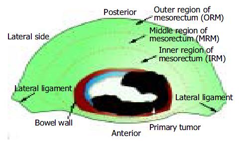
Figure 1 Illustration for regions of mesorectum.
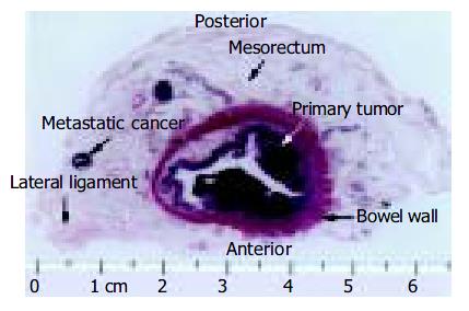
Figure 2 Transverse whole-mount sections of the specimen, HE staining, macroscopic view.
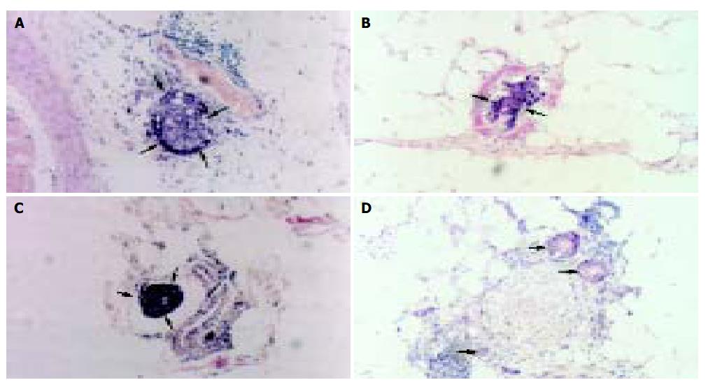
Figure 3 Spread types of microscopic tumor nodules (→) in the mesorectum, HE, × 100.
A: Discrete microscopic tumor nodules; B: Blood vessel invasion; C: Lymphatic vessel invasion; D: Perineural invasion.
Figure 4 Microscopic tumor nodule spread (→) in outer region of the mesorectum (ORM) (4a), CRM (4b) and DMR(4c) , HE, × 100.
- Citation: Wang Z, Zhou ZG, Wang C, Zhao GP, Chen YD, Gao HK, Zheng XL, Wang R, Chen DY, Liu WP. Microscopic spread of low rectal cancer in regions of mesorectum: Pathologic assessment with whole-mount sections. World J Gastroenterol 2004; 10(20): 2949-2953
- URL: https://www.wjgnet.com/1007-9327/full/v10/i20/2949.htm
- DOI: https://dx.doi.org/10.3748/wjg.v10.i20.2949


