Copyright
©The Author(s) 2004.
World J Gastroenterol. Oct 1, 2004; 10(19): 2850-2853
Published online Oct 1, 2004. doi: 10.3748/wjg.v10.i19.2850
Published online Oct 1, 2004. doi: 10.3748/wjg.v10.i19.2850
Figure 1 Submucosal tumors on gastric fundus diagnosed in preoperative examinations.
A: A lesion near to ECJ with a clear-cut margin shown in barium swallow examination of upper gastrointestine; B: A lesion with a clear-cut margin protruded into gastric lumen shown in ultrasonic gastroscopy; C: A hemisphere-like projection in the posterior wall of gastric fundus and an ulcer in its center shown in gastroscopy.
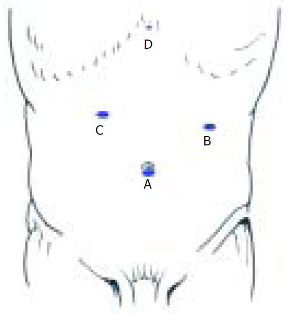
Figure 2 Trocar positions for laparoscopically extraluminal resection of gastric fundus.
A: Endoscope portal (10 mm); B, C: Main working portals (10-12 mm); D: Assisting working por-tal (5 mm).
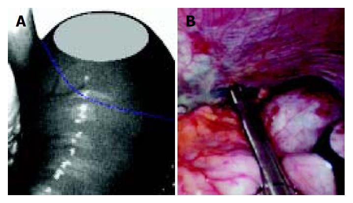
Figure 3 Resection of gastric fundus with Endo GIA.
A: The blue line represents the Endo GIA resection line; B: When Endo GIA was placed near the cardia, special care was taken to ensure that ECJ was not involved.
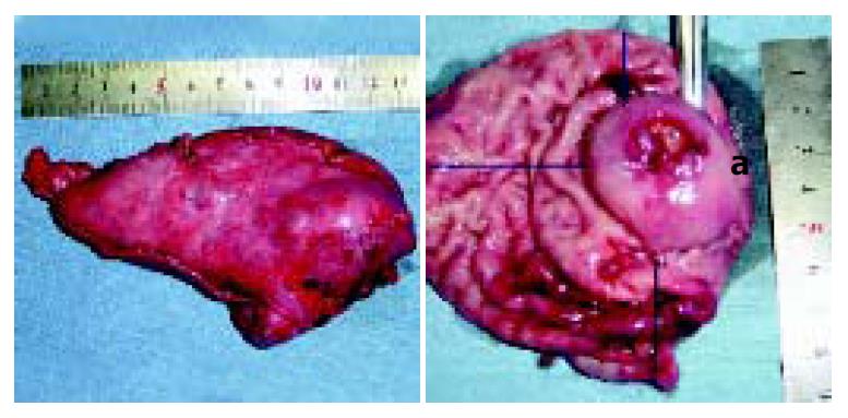
Figure 4 Resected specimens.
Line a: the distance of the tumor to ECJ.
- Citation: Ke ZW, Zheng CZ, Hu MG, Chen DL. Laparoscopic resection of submucosal tumor on posterior wall of gastric fundus. World J Gastroenterol 2004; 10(19): 2850-2853
- URL: https://www.wjgnet.com/1007-9327/full/v10/i19/2850.htm
- DOI: https://dx.doi.org/10.3748/wjg.v10.i19.2850


