Copyright
©The Author(s) 2004.
World J Gastroenterol. Sep 15, 2004; 10(18): 2675-2679
Published online Sep 15, 2004. doi: 10.3748/wjg.v10.i18.2675
Published online Sep 15, 2004. doi: 10.3748/wjg.v10.i18.2675
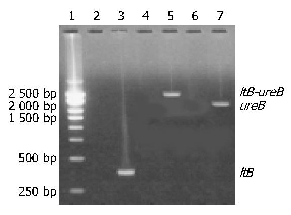
Figure 1 Target amplification fragments of ltB and ureB genes and ltB-ureB fusion gene.
Lane 1: 250 bp DNA marker (BBST); Lanes 2, 4 and 6: Blank controls; Lanes 3 and 5: Target amplification fragments of ltB gene and ltB-ureB fusion gene, respectively; Lane 7: Target recovered fragment of ureB gene from pUCm-T-ureB after digestion with both EcoR V and Xho I.
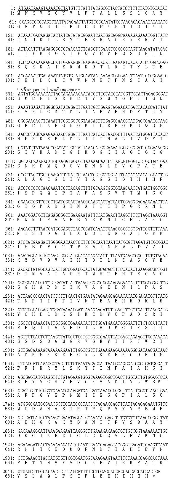
Figure 2 Nucleotide and putative amino acid sequences of ltB-ureB fusion gene.
Note: Underlined areas are sense, linking and antisense primers, respectively. The framed area is the sequence from plasmid pET32a. “*” means stop codon.
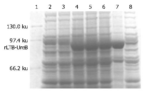
Figure 3 rLTB-UreB expression induced with different dos-ages of IPTG.
Lane 1: Protein marker (Shanghai Shisheng); Lane 2: Blank control; Lane 3: Non-induced with IPTG; Lanes 4-6: Induced with 0.1, 0.5 and 1.0 mmol/L IPTG, respectively; Lanes 7 and 8: Bacterial precipitate and supernatant induced with 0.5 mmol/L IPTG, respectively.
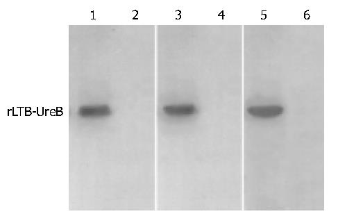
Figure 4 Western blotting of rLTB-UreB with rabbit antibody against whole cell of H pylori and rUreB- and rLTB-UreB-im-munized rabbit antisera.
Lanes 1, 3 and 5: rLTB-UreB with rabbit antibody against whole cell of H pylori, and rUreB- and rLTB-UreB-immunized rabbit antisera, respectively; Lanes 2, 4 and 6: Corresponding blank controls.
-
Citation: Yan J, Wang Y, Shao SH, Mao YF, Li HW, Luo YH. Construction of prokaryotic expression system of
ltB-ureB fusion gene and identification of the recombinant protein immunity and adjuvanticity. World J Gastroenterol 2004; 10(18): 2675-2679 - URL: https://www.wjgnet.com/1007-9327/full/v10/i18/2675.htm
- DOI: https://dx.doi.org/10.3748/wjg.v10.i18.2675


