Copyright
©The Author(s) 2004.
World J Gastroenterol. Sep 1, 2004; 10(17): 2605-2606
Published online Sep 1, 2004. doi: 10.3748/wjg.v10.i17.2605
Published online Sep 1, 2004. doi: 10.3748/wjg.v10.i17.2605
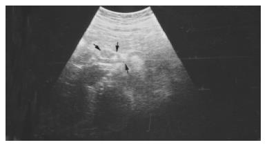
Figure 1 Sonogram showing a fistula (thin arrow) between superior mesenteric artery (arrowhead) and common hepatic artery (thick arrow).
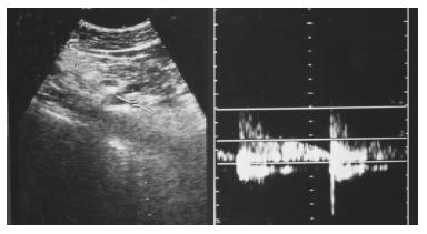
Figure 2 Color doppler sonogram of fistula revealing arterial flow.
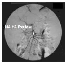
Figure 3 Selective superior mesenteric artery angiography showing a fistula between superior mesenteric artery and common hepatic artery.
- Citation: Kayacetin E, Karaköse S, Karabacakoglu A, Emlik D. Mesenteric Ischemia: An unusual presentation of fistula between superior mesenteric artery and common hepatic artery. World J Gastroenterol 2004; 10(17): 2605-2606
- URL: https://www.wjgnet.com/1007-9327/full/v10/i17/2605.htm
- DOI: https://dx.doi.org/10.3748/wjg.v10.i17.2605


