Copyright
©The Author(s) 2004.
World J Gastroenterol. Aug 15, 2004; 10(16): 2459-2460
Published online Aug 15, 2004. doi: 10.3748/wjg.v10.i16.2459
Published online Aug 15, 2004. doi: 10.3748/wjg.v10.i16.2459
Figure 1 Axial (A) coronal (B) sagittal (C) MIP multislice CT images from the arterial phase showing right subclavian artery originating from the posterior wall of aortic arch, and compression with antreo-right lateral deplacement of esophagus by this right subclavian artery.
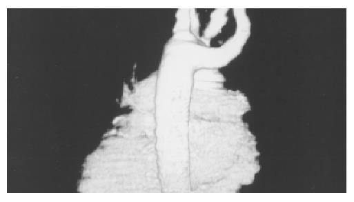
Figure 2 Volume-rendered three-dimensional image by com-puted tomography demonstrating aberrant artery right sub-clavian artery originating from the posterior wall of aortic arch.
- Citation: Kantarceken B, Bulbuloglu E, Yuksel M, Cetinkaya A. Dysphagia lusorium in elderly: A case report. World J Gastroenterol 2004; 10(16): 2459-2460
- URL: https://www.wjgnet.com/1007-9327/full/v10/i16/2459.htm
- DOI: https://dx.doi.org/10.3748/wjg.v10.i16.2459


