Copyright
©The Author(s) 2004.
World J Gastroenterol. Aug 15, 2004; 10(16): 2352-2356
Published online Aug 15, 2004. doi: 10.3748/wjg.v10.i16.2352
Published online Aug 15, 2004. doi: 10.3748/wjg.v10.i16.2352
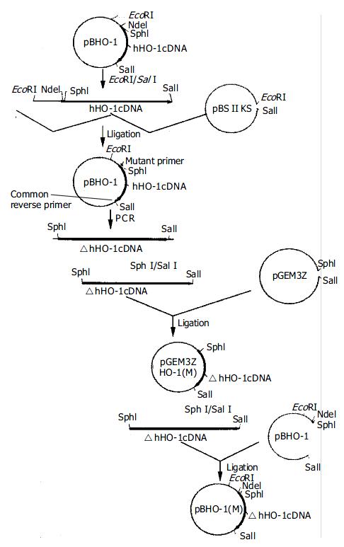
Figure 1 Construction of expression vector pBHO-1(M).
Figure 2 Prime simulated structure of whHO-1 and △hHO-1.
Red: heme; Black: His 25 and Ala 25. Ala 25 loses contacting with heme as His 25 does in the heme-binding pocket.
Figure 3 Changes of bond angle, dihedral angle and chemical bond.
Figure 4 Molecular surfaces and electrostatic potential of whHO-1 and △hHO-1.
The electrostatic potential of active pocket is –1.800.
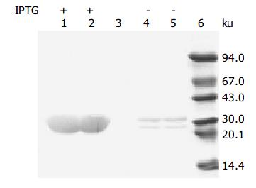
Figure 5 Western blotting of whHO-1 and △hHO-1 expressed in DH5α .
Lanes 1, 2: Expression products of pBHO-1 and pBHO-1(M) in DH5α induced with IPTG; lane 3: Control; lanes 4, 5: Expression products of pBHO-1 and pBHO-1(M) in DH5α not treated with IPTG; lane 6: Marker.
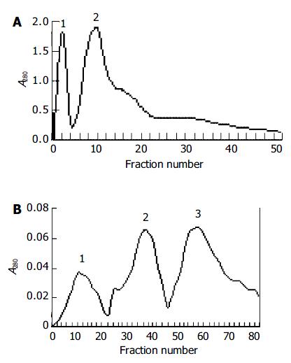
Figure 6 Q-Sepharose Fast Flow column chromatography.
A: pH 7.4 buffer; B: pH 8.4 buffer.
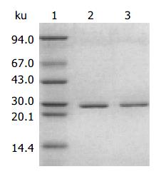
Figure 7 SDS-PAGE analysis of purified whHO-1 and △hHO-1.
Lane 1: Marker; lane 2: whHO-1; lane 3: △hHO-1.
- Citation: Xia ZW, Zhou WP, Cui WJ, Zhang XH, Shen QX, Li YZ, Yu SC. Structure prediction and activity analysis of human heme oxygenase-1 and its mutant. World J Gastroenterol 2004; 10(16): 2352-2356
- URL: https://www.wjgnet.com/1007-9327/full/v10/i16/2352.htm
- DOI: https://dx.doi.org/10.3748/wjg.v10.i16.2352


