Copyright
©The Author(s) 2004.
World J Gastroenterol. Aug 15, 2004; 10(16): 2340-2343
Published online Aug 15, 2004. doi: 10.3748/wjg.v10.i16.2340
Published online Aug 15, 2004. doi: 10.3748/wjg.v10.i16.2340
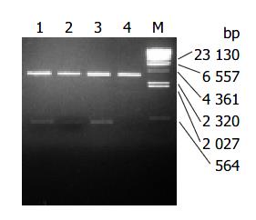
Figure 1 Digestion of recombinant plasmid.
M: λ DNA/ Hind III; Lane 1: pLZ-QV3/ Nco I + Hind III; Lane 2: pLZ-QV2/ Nco I + Hind III; Lane 3: pLZ-QV1/ Nco I + Hind III; Lane 4: pQE-60/ Nco I + Hind III.
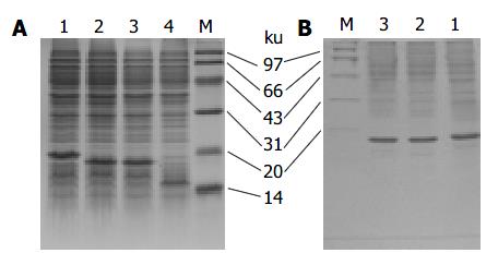
Figure 2 SDS-PAGE of LTB-VacA protein.
M: Marker; Lane 1: LTB-VacA3; Lane 2: LTB-VacA2; Lane 3: LTB-VacA1; Lane 4: LTB. A: LTB-VacA proteins were expressed in the JM109; B: Purification with anti-LTB antibody affinity chromatography.
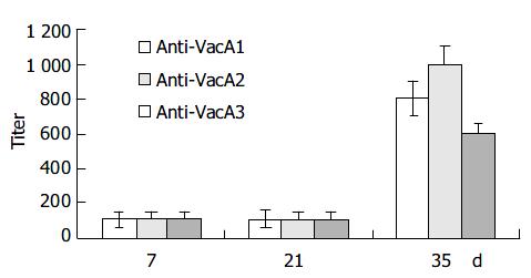
Figure 3 IgG against VacA induced by different LTB-VacA.
Figure 4 Microscopy of large vacuoles induced by VacA in HeLa cells.
A: Normal HeLa cells; B: Vacuolated HeLa cells (Origingal magnification: × 400).
-
Citation: Liu XL, Li SQ, Liu CJ, Tao HX, Zhang ZS. Antigen epitope of
Helicobacter pylori vacuolating cytotoxin A. World J Gastroenterol 2004; 10(16): 2340-2343 - URL: https://www.wjgnet.com/1007-9327/full/v10/i16/2340.htm
- DOI: https://dx.doi.org/10.3748/wjg.v10.i16.2340


