Copyright
©The Author(s) 2004.
World J Gastroenterol. Jul 1, 2004; 10(13): 1943-1947
Published online Jul 1, 2004. doi: 10.3748/wjg.v10.i13.1943
Published online Jul 1, 2004. doi: 10.3748/wjg.v10.i13.1943
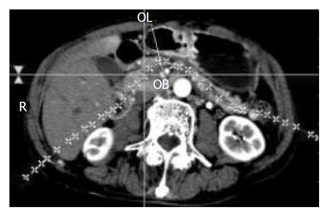
Figure 1 Curve drawn on a transverse image along the pan-creatic duct in a patient with pancreatic adenocarcinoma.
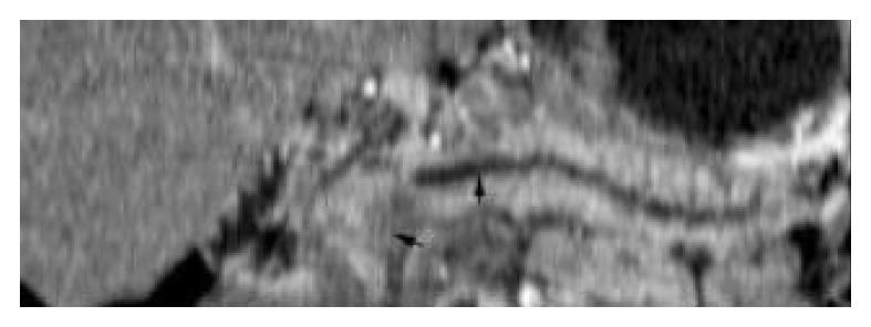
Figure 2 Curve obtained from the curved line on Figure 1 dis-played the hypoattenuation tumor at the head of pancreas and the distal dilated pancreatic duct (arrow).
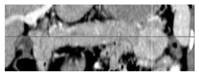
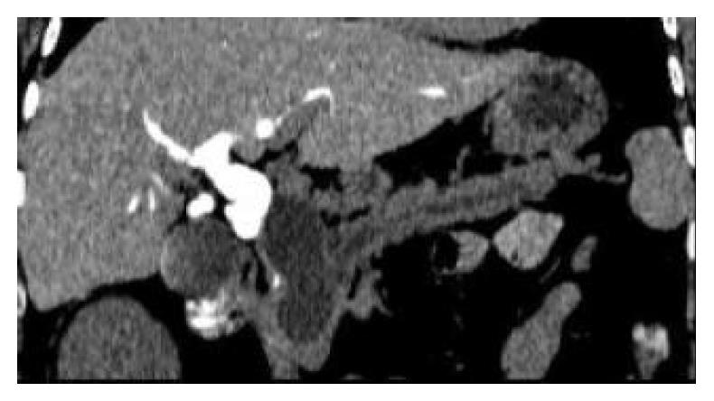
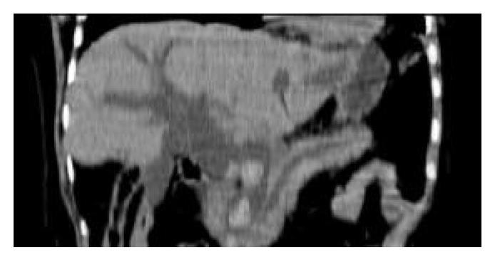
Figure 3 Neuroendocrine tumor of pancreas.
Curved planar reformations during pancreatic parenchyma phase demon-strated the enhanced mass located in the tail of pancreas.
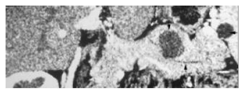
Figure 4 Neuroendocrine tumor of pancreas and peripancreatic pseudocyst.
Curved plane showed a non-enhanced hypoattenuation mass located in the pancreatic parenchyma (arrow) and a pseudocyst at the port of spleen during the pan-creatic parenchyma phase. The normal pancreatic duct was also well depicted (long arrow).
Figure 5 Real cyst of pancreas.
Curved planar reformations obtained after CT cholangiography showed the dilatation and obstruction of bile duct system, and the beaded-like dilatation of pancreatic duct. The cyst located beside the common bile duct. There was no contrast media entrancing into cyst.
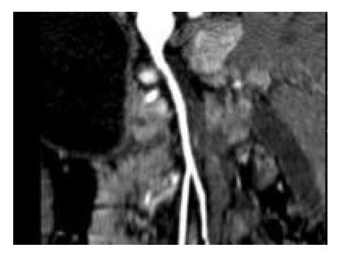
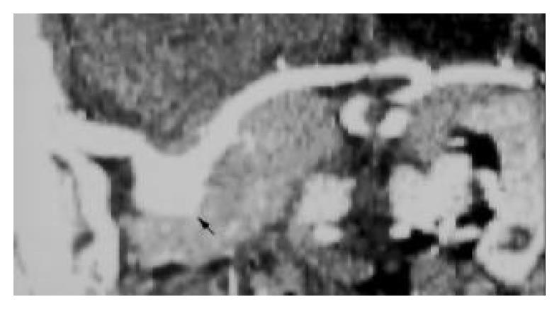
Figure 7 A curved plane tracing the celiac trunk and the splenic artery of a patient with pancreatic adenocarcinoma demon-strated the irregular stricture (arrow).
Figure 8 Ampullary carcinoma.
Curved planar reformations depicted a low attenuation mass around the pancreatic head and the dilatation of both bile duct (arrow) and pancreatic duct (long arrow).
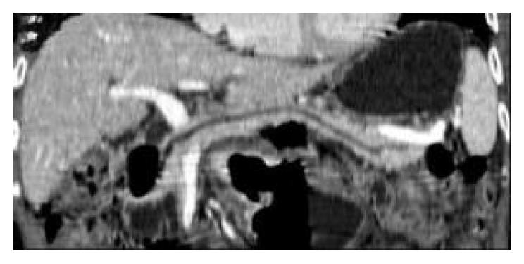
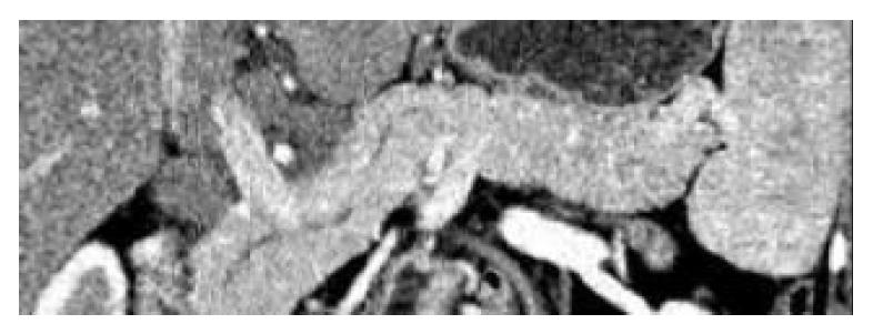
Figure 9 Gastric cancer with peripancreatic adenopathy.
Curved planar reformations depicted the dilatation of the pancreatic duct.
Figure 10 Choledocholithiasis of common bile duct.
Curved plane displayed the dilatation of the bile duct system and the pancreatic duct. The location of stones was depicted clearly.
Figure 11 Peripancreatic adenopathy.
Curved planar refor-mations showed several enlarged lymph nodes located at the pancreatic head.
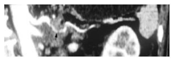
Figure 12 Splenic artery aneurysm.
Curved planar reforma-tions depicted the local dilatation of splenic artery (arrow).
- Citation: Gong JS, Xu JM. Role of curved planar reformations using multidetector spiral CT in diagnosis of pancreatic and peripancreatic diseases. World J Gastroenterol 2004; 10(13): 1943-1947
- URL: https://www.wjgnet.com/1007-9327/full/v10/i13/1943.htm
- DOI: https://dx.doi.org/10.3748/wjg.v10.i13.1943


