Copyright
©The Author(s) 2004.
World J Gastroenterol. Jun 15, 2004; 10(12): 1716-1721
Published online Jun 15, 2004. doi: 10.3748/wjg.v10.i12.1716
Published online Jun 15, 2004. doi: 10.3748/wjg.v10.i12.1716
Figure 1 Parts of DDRT gels including cDNA fragments that show transcriptional alteration in comparing normal esophageal and ESCC tissues (have been pointed with arrows (panel A)), and the results of slot blots containing two microgram RNAs of normal (N) and tumor (T) tissues of esophagus in hybridization with radio-labeled related cDNAs (panel B).
All of the slots have been prepared simultaneously and from the same RNA stocks of T and N. One piece of the slots has been hybridized with radio-labeled GADPH probe as shown in the right most side in panel B.
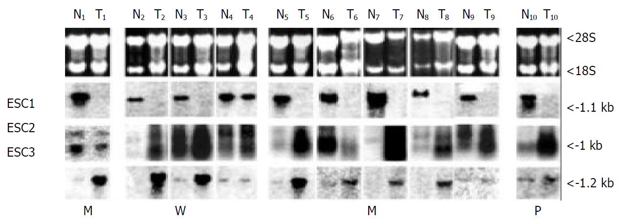
Figure 2 Northern results of patient 1 (RP) and other patients in hybridization with radio-labeled probes of ESC1, ESC2 and ESC3.
Each northern membrane contains about 30 micrograms RNA of normal (N) and tumor (T) tissues of esophagus. The differentiation status of each tumor has been shown as W: Well differentiated tumor; M: Moderately differentiated tumor; P: poorly differentiated tumor. The estimated sizes of each bands has been shown on the right side and the ethidium bromide stained gel on top shows the relative amounts of RNAs loaded in each lane.
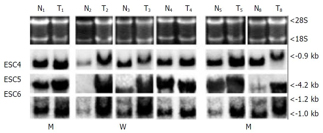
Figure 3 Northern results of patient 1 (RP) and other patients in hybridization with radio-labeled probes of ESC4, ESC5 and ESC6.
Each northern membrane contains about 20 μg RNA of normal (N) and tumor (T) tissues of esophagus. The differentiation status of each tumor has been shown as W: Well differentiated tumor; M: Moderately differentiated tumor. The estimated sizes of each bands has been shown on the right side and the ethidium bromide stained gel on top shows the relative amounts of RNAs loaded in each lane.
- Citation: Kazemi-Noureini S, Colonna-Romano S, Ziaee AA, Malboobi MA, Yazdanbod M, Setayeshgar P, Maresca B. Differential gene expression between squamous cell carcinoma of esophageus and its normal epithelium; altered pattern of mal, akr1c2, and rab11a expression. World J Gastroenterol 2004; 10(12): 1716-1721
- URL: https://www.wjgnet.com/1007-9327/full/v10/i12/1716.htm
- DOI: https://dx.doi.org/10.3748/wjg.v10.i12.1716


