Copyright
©The Author(s) 2004.
World J Gastroenterol. Jun 1, 2004; 10(11): 1639-1642
Published online Jun 1, 2004. doi: 10.3748/wjg.v10.i11.1639
Published online Jun 1, 2004. doi: 10.3748/wjg.v10.i11.1639
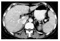
Figure 1 Serohepatic type: multiple-nodular hypodense lesions in the subcapsule of liver and thickened subcapsule of quadrotus lobe on CT.
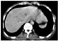
Figure 2 Miliary tuberculosis: scattered distribution of multiple, miliary, micronodular and low-density lesions in liver.
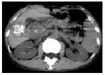
Figure 3 Nodular tuberculosis: singular low-density mass with multiple flecked calcifications in the right lobe of liver and tuberculous lymphadenopathy encroaching on head of pancreas.
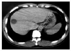
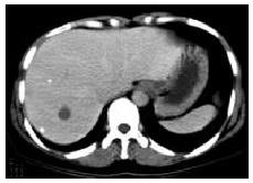
Figure 4 Nodular tuberculosis: cystic mass with 23 Hu in the right lobe of liver.
Figure 5 Mixed tuberculosis: singular, round-like and low-density lesion and multiple miliary calcifications in the right lobe of liver.
Figure 6 MR T2-weighted images showing singular, round-like hypointense lesion.
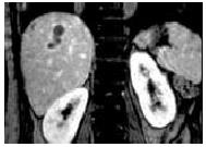
Figure 7 MRI showing irregular strip lesion with multilocular enhancement on coronary plane.
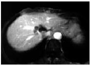
Figure 8 Enhanced MRI showing multiple micronodular lesions fusing into multiloculated cystic mass near the second porta hepatis.
- Citation: Yu RS, Zhang SZ, Wu JJ, Li RF. Imaging diagnosis of 12 patients with hepatic tuberculosis. World J Gastroenterol 2004; 10(11): 1639-1642
- URL: https://www.wjgnet.com/1007-9327/full/v10/i11/1639.htm
- DOI: https://dx.doi.org/10.3748/wjg.v10.i11.1639


