Copyright
©The Author(s) 2004.
World J Gastroenterol. Jun 1, 2004; 10(11): 1634-1638
Published online Jun 1, 2004. doi: 10.3748/wjg.v10.i11.1634
Published online Jun 1, 2004. doi: 10.3748/wjg.v10.i11.1634
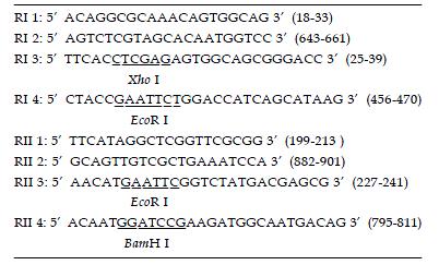
Figure 1 Primer pairs for PCR reactions.
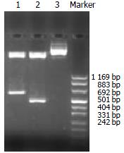
Figure 2 Enzyme-cutting identification of the recombinant plasmids, Lane 1: pANTI-RII, Lane 2: pANTI-RI, Lane 3: pcDNA3.
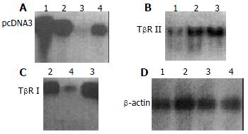
Figure 3 Expression of exogenous transfected gene assessed by Northern blot, A: Hybridization using 32P labeled oligo-nucleotide probe which targets T7 promoter; B: Hybridization by using 32P labeled TβR II cDNA probe; C: Hybridization by using 32P labeled TβR I cDNA probe; D: β-actin probe used as control.
Lane 1: Antisense TβR II group; Lane 2: pcDNA3 con-trol group; Lane 3: Disease control group; Lane 4: Antisense TβR I group.
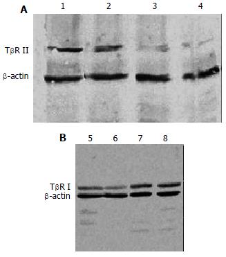
Figure 4 Hepatic protein expression of TβR I, TβR II assessed by Western blot, A: Hepatic protein expression of TβR II; B: Hepatic protein expression of TβR I; Lanes 1,8: Disease control group; Lanes 2,7: pcDNA3 control group; Lane 3: Antisense TβR II group; Lanes 4,6: Normal control group; Lane 5: Antisense TβR I group.
- Citation: Jiang W, Yang CQ, Liu WB, Wang YQ, He BM, Wang JY. Blockage of transforming growth factor β receptors prevents progression of pig serum-induced rat liver fibrosis. World J Gastroenterol 2004; 10(11): 1634-1638
- URL: https://www.wjgnet.com/1007-9327/full/v10/i11/1634.htm
- DOI: https://dx.doi.org/10.3748/wjg.v10.i11.1634


