Copyright
©The Author(s) 1995.
World J Gastroenterol. Oct 1, 1995; 1(1): 18-20
Published online Oct 1, 1995. doi: 10.3748/wjg.v1.i1.18
Published online Oct 1, 1995. doi: 10.3748/wjg.v1.i1.18
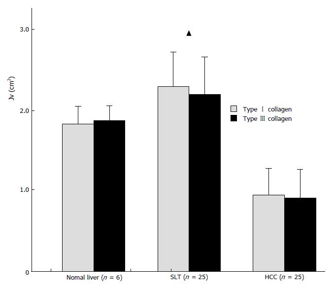
Figure 1 Content of types I and III collagen in HCC.
HCC vs normal liver and vs SLT, P < 0.01; SLT vs normal liver, P < 0.05. HCC: Hepatocellular carcinoma; SLT: Surrounding liver tissue.
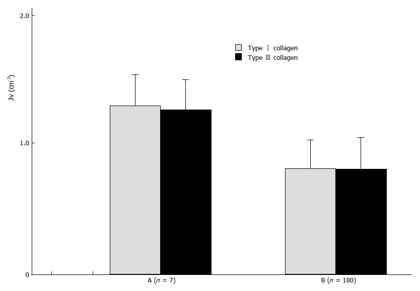
Figure 2 Content of types I and III collagen in HCC with invasion and/or metastasis (B) and in HCC without invasion and/or metastasis (A).
A vs B, P < 0.01. HCC: Hepatocellular carcinoma.
- Citation: Wang YJ, Sun ZQ, Yu JJ, Xu XZ, Zhang X, Quan QZ. Biological effects of types I and III collagens in human hepatocellular carcinoma tissue. World J Gastroenterol 1995; 1(1): 18-20
- URL: https://www.wjgnet.com/1007-9327/full/v1/i1/18.htm
- DOI: https://dx.doi.org/10.3748/wjg.v1.i1.18


