Copyright
©The Author(s) 2025.
Artif Intell Gastrointest Endosc. Mar 28, 2025; 6(1): 105674
Published online Mar 28, 2025. doi: 10.37126/aige.v6.i1.105674
Published online Mar 28, 2025. doi: 10.37126/aige.v6.i1.105674
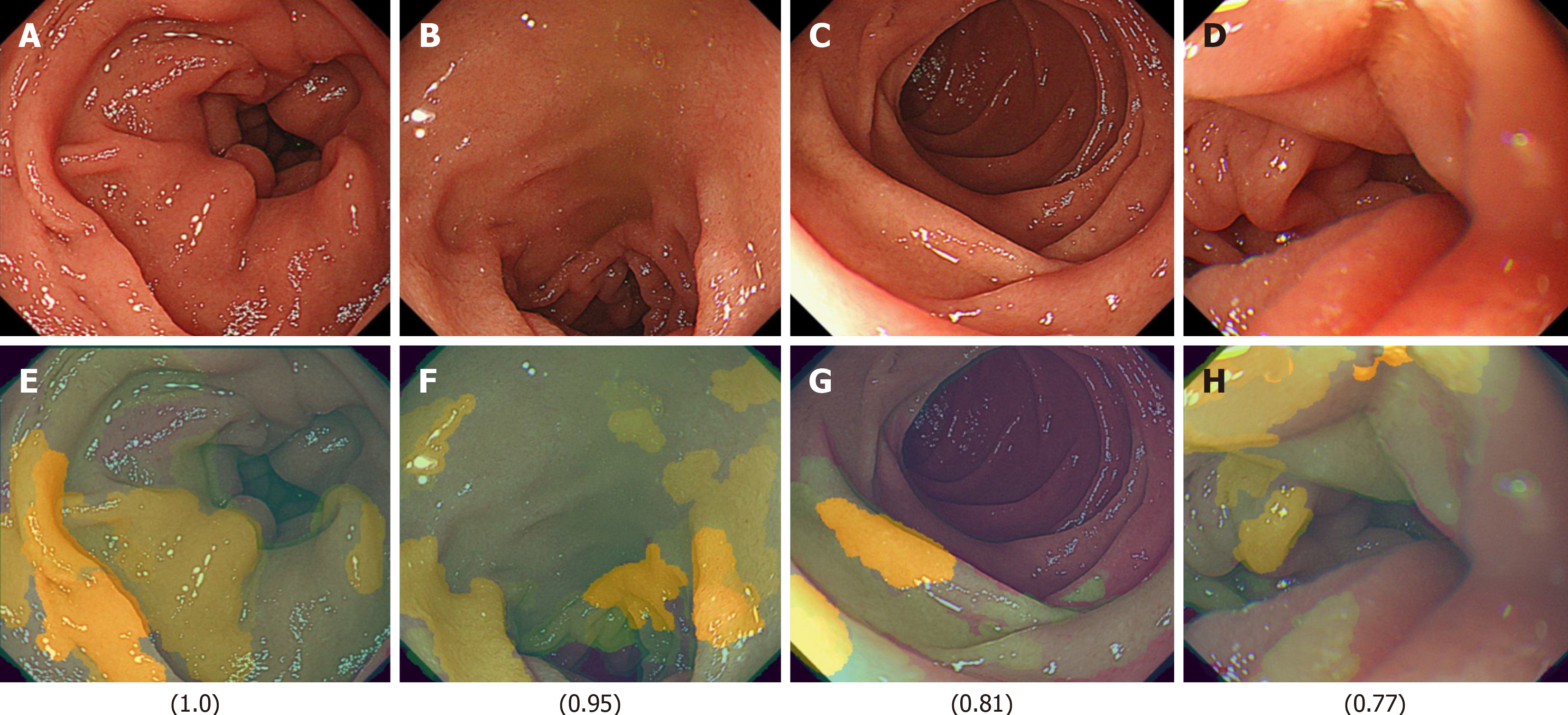
Figure 6 Representative light images and their corresponding XAI (XRAI) images with high confidence scores (ranging from 0 to 1) in the duodenal image artificial intelligence model for detecting the presence of functional dyspepsia in Helicobacter pylori-infected patients.
The values displayed correspond to the assigned scores. A: A light image of the duodenal mucosa with a high artificial intelligence (AI) confidence score of 1.0, indicating a strong prediction of functional dyspepsia; B: Another light image with a high AI confidence score of 0.95; C: A light image with a score of 0.81; D: A light image with a score of 0.77; E: The XAI (XRAI) visualization corresponding to A, highlighting the regions the AI model identified as most influential in its prediction; F: The XAI (XRAI) visualization corresponding to B; G: The XAI (XRAI) visualization corresponding to C; H: The XAI (XRAI) visualization corresponding to D.
- Citation: Mihara H, Nanjo S, Motoo I, Ando T, Fujinami H, Yasuda I. Artificial intelligence model on images of functional dyspepsia. Artif Intell Gastrointest Endosc 2025; 6(1): 105674
- URL: https://www.wjgnet.com/2689-7164/full/v6/i1/105674.htm
- DOI: https://dx.doi.org/10.37126/aige.v6.i1.105674


