Copyright
©The Author(s) 2025.
Artif Intell Gastrointest Endosc. Mar 28, 2025; 6(1): 105674
Published online Mar 28, 2025. doi: 10.37126/aige.v6.i1.105674
Published online Mar 28, 2025. doi: 10.37126/aige.v6.i1.105674
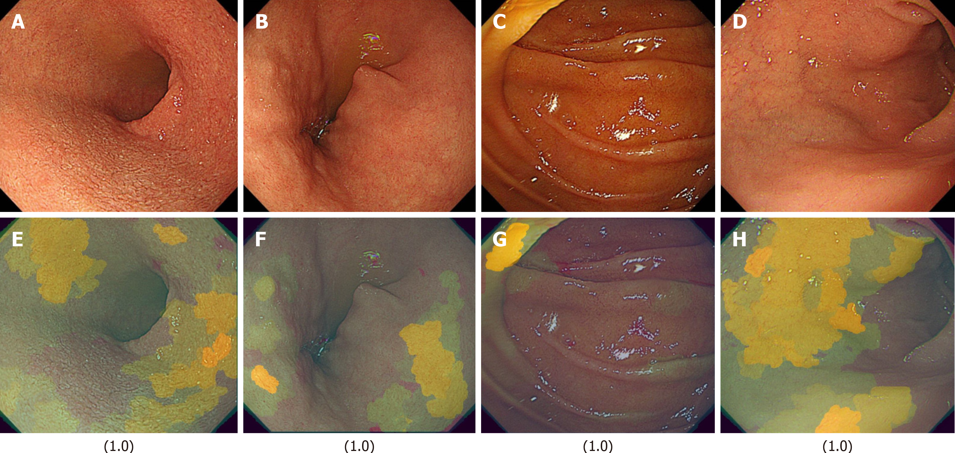
Figure 5 Representative light images and their corresponding XAI (XRAI) images with high confidence scores (1.
0) in the duodenal image artificial intelligence model for detecting the absence of functional dyspepsia in Helicobacter pylori-infected patients. The values displayed correspond to the assigned scores. A: A light image of the duodenal mucosa with a confidence score of 1.0, indicating a strong prediction of the absence of functional dyspepsia; B: Another light image with a score of 1.0; C: A light image with a confidence score of 1.0; D: A light image with a confidence score of 1.0; E: The XAI (XRAI) visualization corresponding to A, highlighting the regions the artificial intelligence model considered important for its classification; F: The XAI (XRAI) visualization corresponding to B; G: The XAI (XRAI) visualization corresponding to C; H: The XAI (XRAI) visualization corresponding to D.
- Citation: Mihara H, Nanjo S, Motoo I, Ando T, Fujinami H, Yasuda I. Artificial intelligence model on images of functional dyspepsia. Artif Intell Gastrointest Endosc 2025; 6(1): 105674
- URL: https://www.wjgnet.com/2689-7164/full/v6/i1/105674.htm
- DOI: https://dx.doi.org/10.37126/aige.v6.i1.105674


