Copyright
©The Author(s) 2020.
Artif Intell Med Imaging. Jun 28, 2020; 1(1): 65-69
Published online Jun 28, 2020. doi: 10.35711/aimi.v1.i1.65
Published online Jun 28, 2020. doi: 10.35711/aimi.v1.i1.65
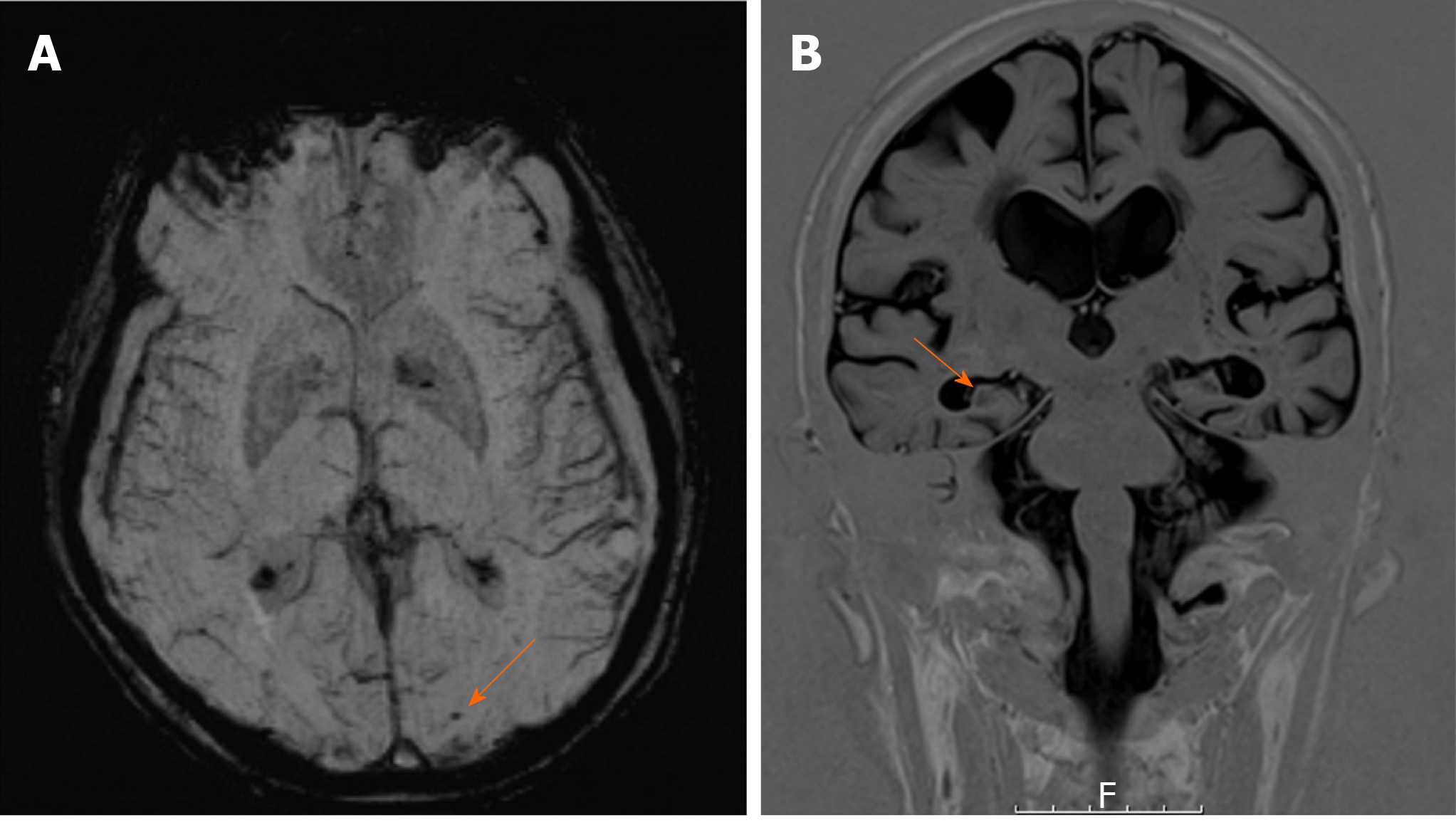
Figure 1 Magnetic resonance imaging of the patient.
A: Susceptibility weighted magnetic resonance imaging (MRI) showing left occipital microhaemorrhage (arrow), suggestive of cerebral amyloid angiopathy; B: Coronal view T1-weighted MRI showing bilateral mesial temporal atrophy (arrow).
- Citation: Arberry J, Singh S, Mizoguchi RA. Cerebral amyloid angiopathy vs Alzheimer’s dementia: Diagnostic conundrum. Artif Intell Med Imaging 2020; 1(1): 65-69
- URL: https://www.wjgnet.com/2644-3260/full/v1/i1/65.htm
- DOI: https://dx.doi.org/10.35711/aimi.v1.i1.65


