Copyright
©The Author(s) 2020.
Artif Intell Med Imaging. Jun 28, 2020; 1(1): 40-49
Published online Jun 28, 2020. doi: 10.35711/aimi.v1.i1.40
Published online Jun 28, 2020. doi: 10.35711/aimi.v1.i1.40
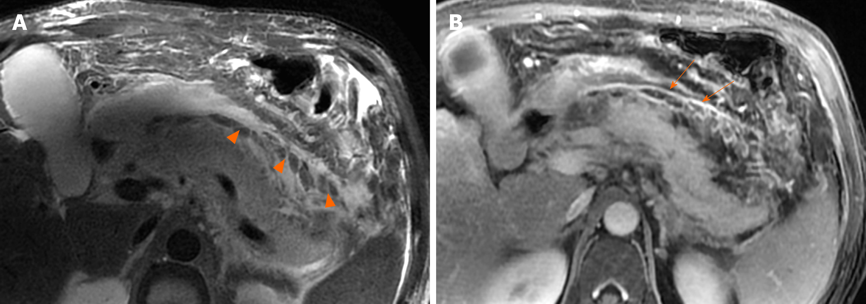
Figure 6 A 46-year-old man with necrotizing pancreatitis.
A: Magnetic resonance imaging axial T2WI image shows a number of patchy, strip-shaped T2-hypointense components (necrotic adipose fragments) (arrowheads) among T2-hyperintense fluid; B: Axial contrast-enhanced magnetic resonance imaging shows viable capsule enhancement (arrows).
- Citation: Xiao B. Acute pancreatitis: A pictorial review of early pancreatic fluid collections. Artif Intell Med Imaging 2020; 1(1): 40-49
- URL: https://www.wjgnet.com/2644-3260/full/v1/i1/40.htm
- DOI: https://dx.doi.org/10.35711/aimi.v1.i1.40


