Copyright
©The Author(s) 2017.
World J Meta-Anal. Dec 26, 2017; 5(6): 132-149
Published online Dec 26, 2017. doi: 10.13105/wjma.v5.i6.132
Published online Dec 26, 2017. doi: 10.13105/wjma.v5.i6.132
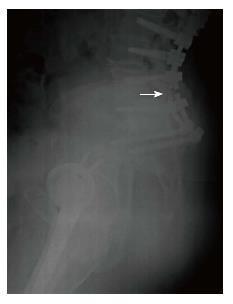
Figure 4 An example of radiographic findings of rod fracture.
Bilateral L5-S1 rod fractures are shown by white arrow; they were diagnosed consequently at 12 and 20 mo follow-up after surgical correction of adult spine deformity, and accompanied with L5-S1 pseudo-arthrosis. The pseudo-arthrosis at L5-S1 was diagnosed simultaneously with the second rod fracture at 20 mo follow-up. The patient experienced increasing low back pain and sagittal imbalance. A revision operation was performed at 21 mo follow-up with revision of the fusion and an osteotomy to correct residual sagittal imbalance.
- Citation: Barton C, Noshchenko A, Patel VV, Cain CMJ, Kleck C, Burger EL. Different types of mechanical complications after surgical correction of adult spine deformity with osteotomy. World J Meta-Anal 2017; 5(6): 132-149
- URL: https://www.wjgnet.com/2308-3840/full/v5/i6/132.htm
- DOI: https://dx.doi.org/10.13105/wjma.v5.i6.132


